Comprehensive immune checkpoint research requires integrated analysis of checkpoint receptor signaling, T-cell activation and exhaustion states, tumor immune microenvironment composition, antigen presentation mechanisms, and functional immune responses to define mechanisms of immune escape and therapeutic sensitivity.
We provide an integrated research solution — from checkpoint signaling targets to immune functional assays — supporting studies of immune regulation and cancer immunotherapy.
## Key Target Highlights
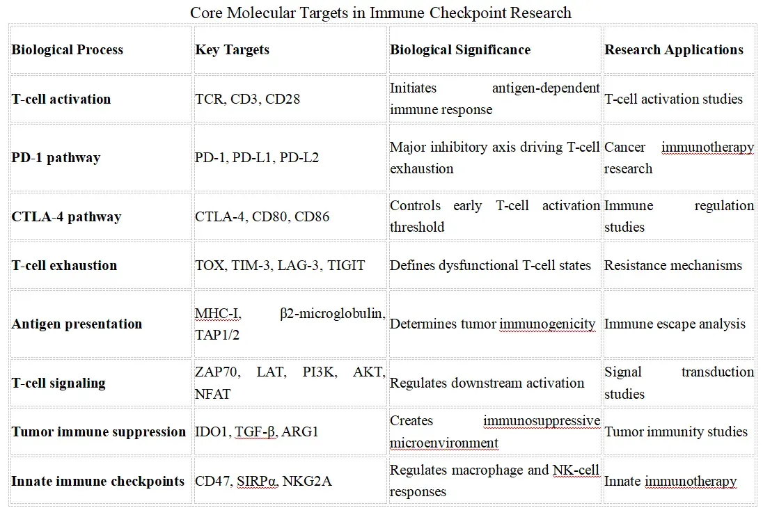
### Key research trend:
Modern immune checkpoint research is moving beyond individual inhibitory receptors toward a systems-level understanding of immune cell states, tumor microenvironment interactions, antigen presentation, metabolic regulation, and combination therapeutic strategies. Current studies increasingly integrate single-cell sequencing, spatial profiling, and functional immune assays to identify mechanisms of response and resistance to checkpoint blockade therapies.
## Recommended Immune Checkpoint Marker Strategy
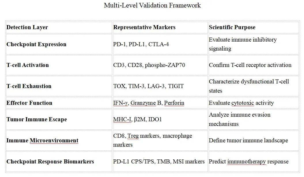
## Core Immune Checkpoint Validation Strategy
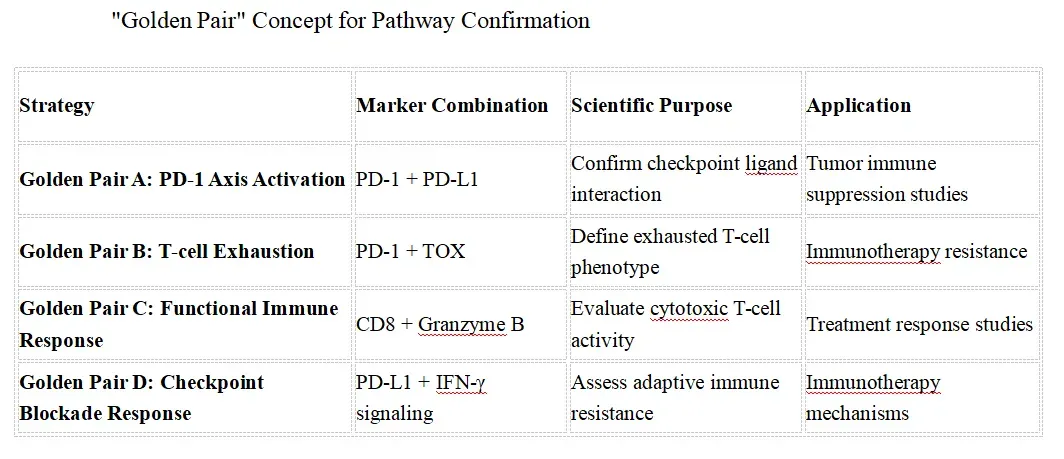
## Competitive Technology Landscape
Immune Checkpoint Research Workflow
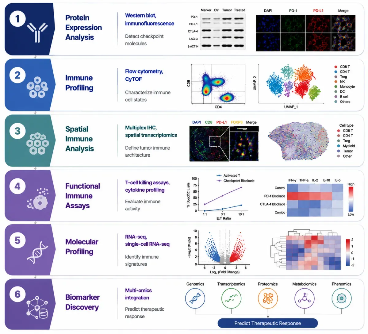
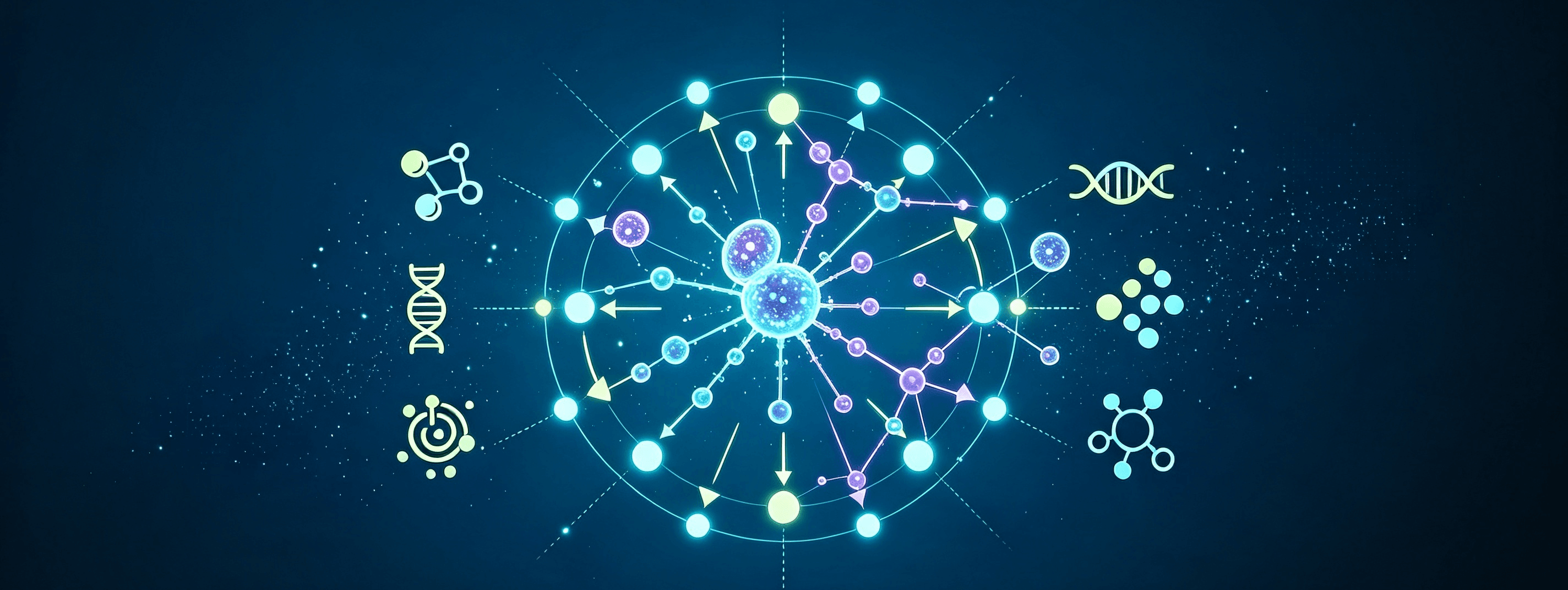
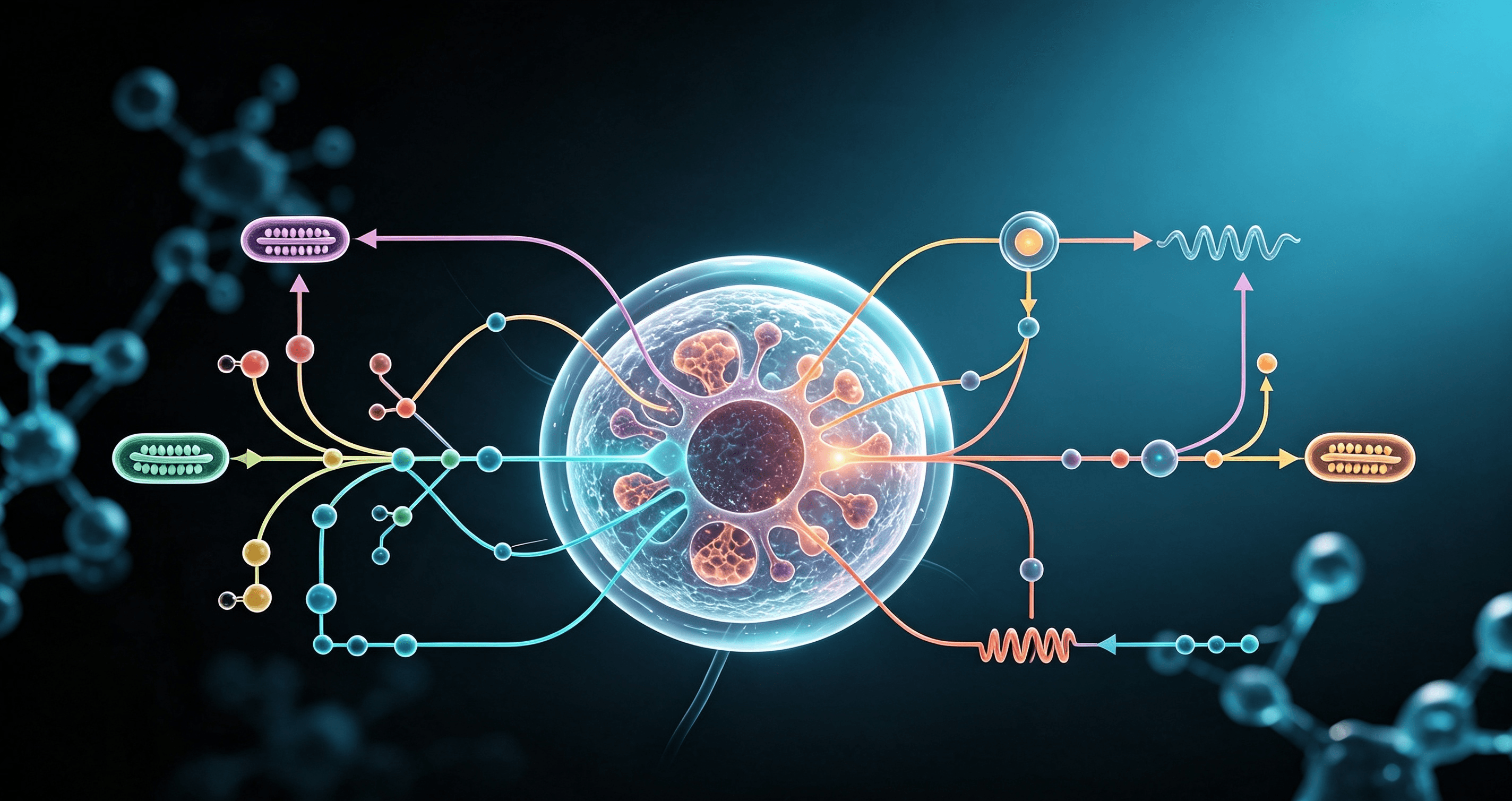
Immune checkpoint research requires an integrated workflow combining protein validation, immune cell profiling, spatial analysis, functional assessment, molecular characterization, and multi-omics integration. This comprehensive approach enables researchers to understand immune regulation mechanisms, define tumor immune landscapes, and identify predictive biomarkers for immunotherapy response.
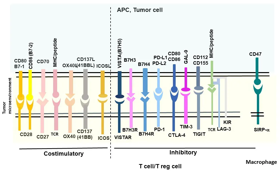
## Pathway Overview
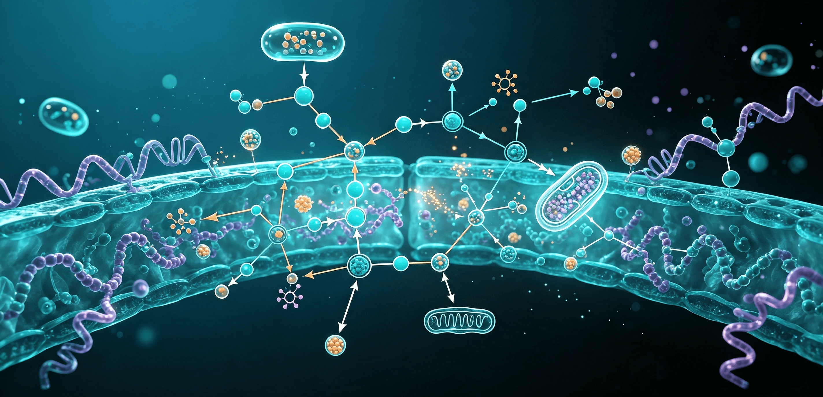
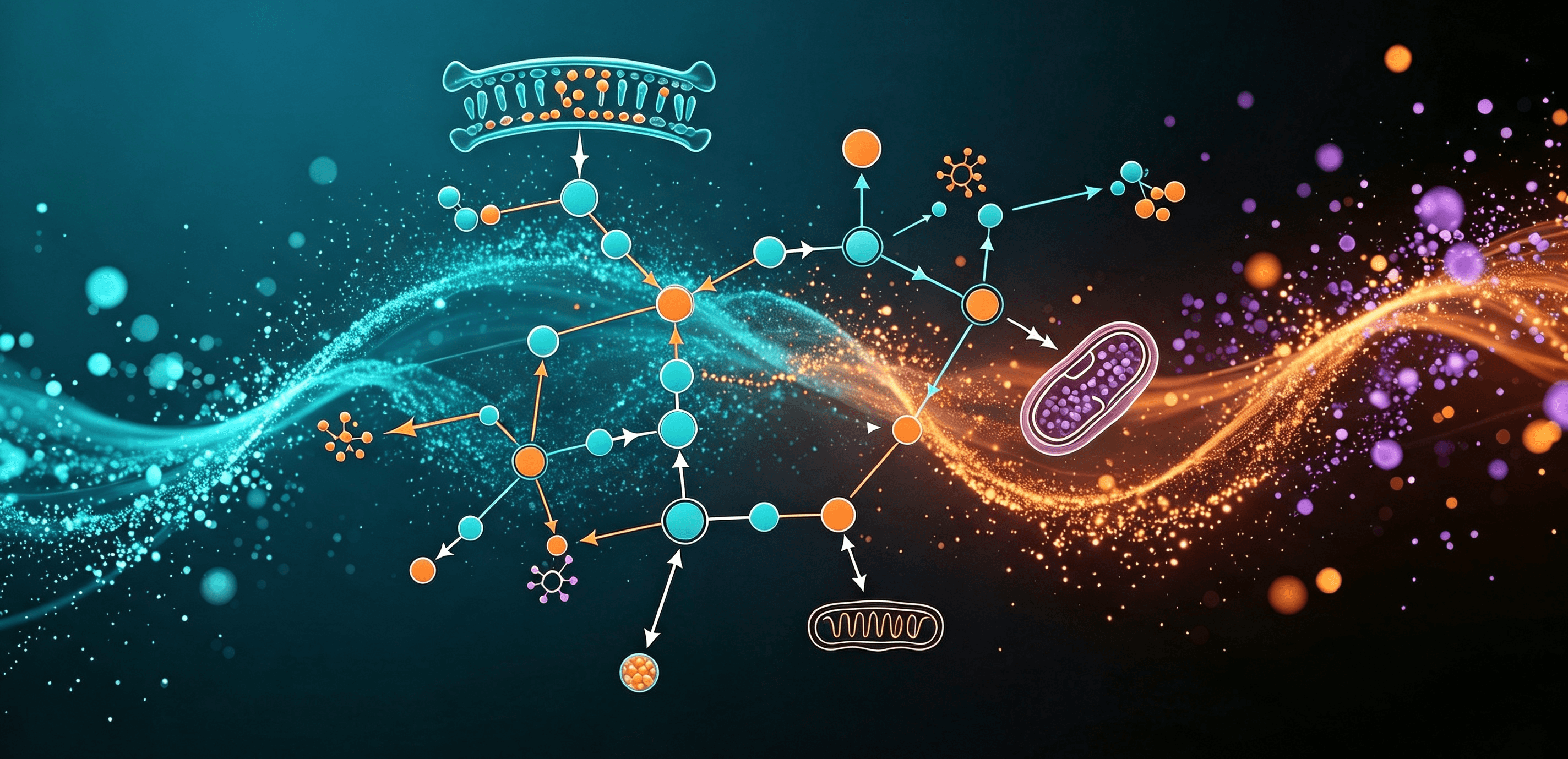
Immune checkpoint pathways maintain immune balance by regulating T-cell activation, tolerance, and immune responses. Tumors exploit inhibitory pathways such as PD-1/PD-L1 and CTLA-4 to suppress anti-tumor immunity and promote immune escape. Emerging checkpoint pathways, including LAG-3, TIM-3, TIGIT, and VISTA, expand the landscape of immune regulation and immunotherapy research.
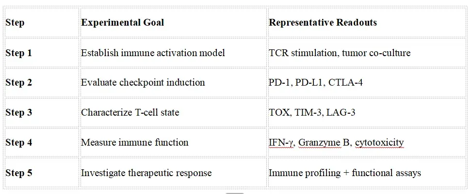
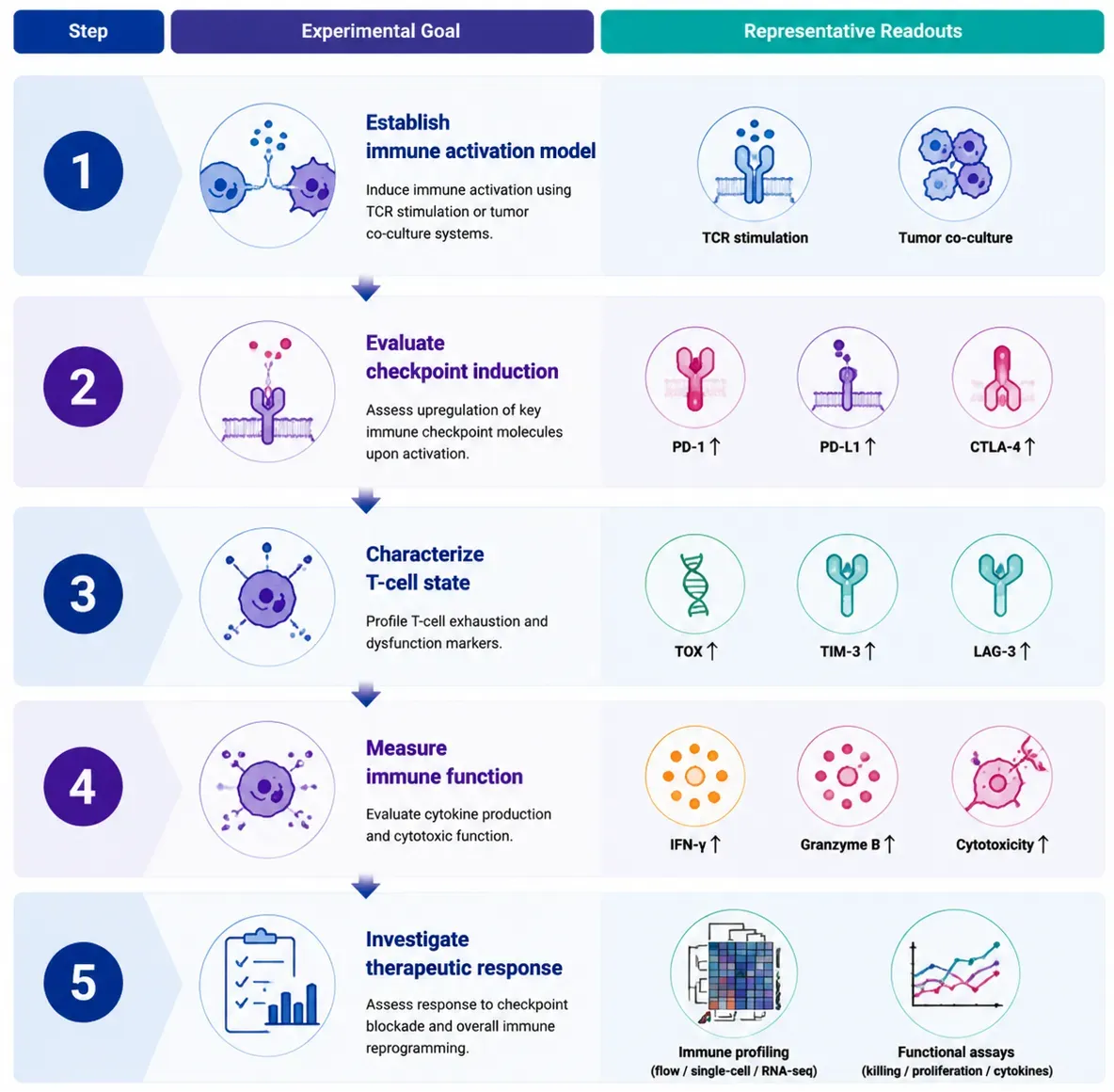
## Recommended Experimental Validation Workflow
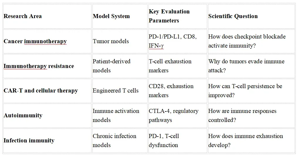
## Featured Research Application Examples
## Frequently Asked Questions
Q1.Which markers should be used to characterize immune checkpoint signaling?
Immune checkpoint analysis requires evaluation of both checkpoint molecules and downstream immune signaling pathways. Common checkpoint markers include PD-1 (PDCD1), PD-L1 (CD274), CTLA-4, LAG-3, TIM-3, and TIGIT. Additional markers such as CD3, CD8, CD4, FOXP3, and Granzyme B help define immune cell populations and functional states. Downstream pathway analysis often includes TCR signaling, NFAT, NF-κB, PI3K/AKT, and MAPK pathways to understand checkpoint-mediated immune regulation.
Q2. How can researchers evaluate immune checkpoint activation and immune cell function?
Immune checkpoint studies should combine expression analysis with functional assays. Common approaches include flow cytometry, immunohistochemistry (IHC), immunofluorescence (IF), and Western blotting to assess checkpoint protein expression and localization. Functional evaluation includes T-cell activation assays, cytokine profiling (IFN-γ, IL-2, TNF-α), cytotoxicity assays, and co-culture systems to determine how checkpoint signaling affects immune responses.
Q3. . What are the major signaling pathways involved in immune checkpoint regulation?
Immune checkpoints regulate T-cell activation through multiple signaling networks. The PD-1/PD-L1 axis suppresses T-cell activity by recruiting phosphatases such as SHP-2, leading to inhibition of TCR/CD28 signaling, PI3K/AKT, and MAPK pathways. The CTLA-4 pathway regulates early T-cell activation by competing with CD28 for B7 ligands. Additional pathways involving LAG-3, TIM-3, TIGIT, and metabolic regulators contribute to immune exhaustion and tumor immune escape.
Q4. What are the common challenges and pitfalls in immune checkpoint research?
Major challenges include immune heterogeneity, dynamic checkpoint expression, species differences, and tumor microenvironment complexity. A common pitfall is interpreting checkpoint expression alone as evidence of immune suppression. Researchers should integrate checkpoint profiling, immune cell characterization, functional validation, and appropriate controls to distinguish immune activation, exhaustion, and therapeutic response mechanisms.
Q5. How can immune checkpoint research be translated into immunotherapy development?
Advanced immune checkpoint studies integrate single-cell sequencing, spatial transcriptomics, multiplex imaging, proteomics, and biomarker analysis to identify immune states and predict therapeutic responses. These approaches support discovery of immune checkpoint biomarkers, combination therapy strategies, and mechanisms of resistance to immune checkpoint inhibitors in cancer and other immune-related diseases.
## Key References
1.Roerden M., Spranger S. (2025).Cancer immune evasion, immunoediting and intratumour heterogeneity.Nature Reviews Immunology. 25:353–369.
2.Zhang Z., Zhang C. (2025).Regulation of cGAS–STING signalling and its diversity of cellular outcomes.Nature Reviews Immunology. 25:425–444.
3.Lotze M.T., Olejniczak S.H., Skokos D. (2024).CD28 co-stimulation: novel insights and applications in cancer immunotherapy.Nature Reviews Immunology. 24(12):878–895.
4.Butterfield L.H., Najjar Y.G. (2024).Immunotherapy combination approaches: mechanisms, biomarkers and clinical observations.Nature Reviews Immunology. 24(6):399–416.
5. Schenkel J.M., Pauken K.E. (2023).Localization, tissue biology and T cell state — implications for cancer immunotherapy.Nature Reviews Immunology. 23:807–823.
6.Hato S.V., Khong A., Fiechter M., et al. (2024).Immune checkpoint blockade: current progress and future directions.Nature Reviews Cancer. 24:585–605.
7.Wei S.C., Duffy C.R., Allison J.P. (2024).Fundamental mechanisms of immune checkpoint blockade therapy.Cancer Discovery. 14:1120–1137.
8.Chauvin J.M., Pagliano O., Fourcade J., et al. (2022).TIGIT and the tumor microenvironment: emerging mechanisms and therapeutic opportunities.Trends in Cancer. 8:914–928.
Explore Resources












 Search Products by Molecule Initial