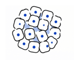
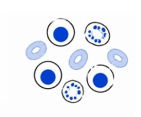
Stem cells are a unique group of cells characterized by their ability to self-renew and possess differentiation potential. Unlike highly differentiated mature cells with fixed functions, stem cells remain in a relatively undifferentiated state and can continuously proliferate to maintain their own cell pool. Under the regulation of external signals, growth factors, and the cellular microenvironment (niche), stem cells can differentiate into specialized cell types with specific functions. Based on their differentiation capacity and origin, stem cells are mainly classified into pluripotent stem cells and adult or tissue-specific stem cells. Due to their remarkable regenerative capacity and developmental potential, stem cells are considered fundamental components responsible for tissue development, homeostasis, and repair following injury.
Explore new frontiers in stem cell research and advance innovation in life sciences.
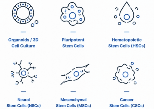
Our stem cell product portfolio provides researchers worldwide with comprehensive culture solutions, supporting the entire workflow from stem cell maintenance, expansion, and differentiation to organoid construction and the development of 3D cell models. Focusing on key research areas including induced pluripotent stem cells (iPSCs), embryonic stem cells (ESCs), mesenchymal stem cells (MSCs), neural stem cells (NSCs), hematopoietic stem cells (HSCs), and cancer stem cells (CSCs), we offer high-performance culture systems and specialized products to help researchers establish cell models that more closely recapitulate human physiological environments.
Through advanced culture technologies and innovative solutions, we empower researchers to gain deeper insights into tissue development, disease mechanisms, cellular functional regulation, and therapeutic responses, providing strong support for regenerative medicine, precision medicine, and drug discovery.
Pluripotent Stem Cells — What Are Embryonic Stem Cells (ESCs) and Induced Pluripotent Stem Cells (iPSCs)?
Currently, human pluripotent stem cells (hPSCs) being explored for clinical applications mainly include two major types: embryonic stem cells (Embryonic Stem Cells, ESCs) and induced pluripotent stem cells (Induced Pluripotent Stem Cells, iPSCs).
Embryonic stem cells (ESCs) and induced pluripotent stem cells (iPSCs) are highly valuable pluripotent stem cell types characterized by their ability to undergo continuous self-renewal and differentiate into cells derived from the three germ layers: ectoderm, mesoderm, and endoderm. These three germ layers give rise to nearly all mature cell types in the human body. Therefore, ESCs and iPSCs have become essential model systems for studying cell fate determination, tissue development, disease mechanisms, and regenerative medicine. ESCs are derived from the inner cell mass of early-stage embryos, whereas iPSCs are generated by reprogramming mature somatic cells into a pluripotent state through the introduction of specific reprogramming factors. Owing to their unlimited expansion capacity and broad differentiation potential, these two cell types are widely applied in disease modeling, drug discovery and safety evaluation, cell therapy development, and tissue engineering research.
Embryonic and Induced Pluripotent Stem Cell Research Workflow
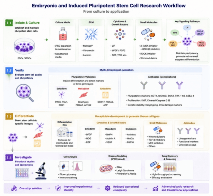
Our embryonic stem cell (ESC) and induced pluripotent stem cell (iPSC) research product portfolio is designed around the complete experimental workflow, providing researchers with an integrated solution covering stem cell establishment, pluripotency validation, directed differentiation, and functional studies. Through high-quality reagents and optimized culture systems that support the entire stem cell research pipeline, we help researchers improve experimental consistency, simplify operational procedures, and accelerate advances in fundamental stem cell research and translational applications.
Isolate & Culture
Supporting the isolation, expansion, and long-term maintenance of pluripotent stem cells, we provide optimized culture media, essential supplements, and specialized culture systems to help maintain an undifferentiated state, promote stable proliferation, and establish high-quality stem cell culture platforms.
- iPSC Expansion and Maintenance Medium
- Conditioned Medium (with cultured cell images displayed)
- Extracellular Matrix (ECM)
- Cytokines (Cytokines for ESC and iPSC Expansion)
- Small Molecules — To maintain long-term expansion and an undifferentiated state of pluripotent stem cells, precise regulation of intracellular and extracellular signaling networks is essential, including key pathways such as Wnt, LIF, FGF/ERK, TGF-β/BMP, and Rho/ROCK signaling. We provide high-quality small molecule modulators and research tools to support ESC/iPSC culture, enabling enhanced proliferation, self-renewal, pluripotency maintenance, and cell fate regulation, helping researchers establish stable, efficient, and reproducible stem cell culture systems.
By precisely regulating critical molecular pathways, these small molecules can be applied to optimize feeder-free culture systems, establish 2i culture conditions, and maintain pluripotent stem cell states. For example, the combination of a MEK inhibitor and a GSK-3β inhibitor in the classical 2i system regulates ERK and Wnt signaling pathways, reduces spontaneous differentiation, and promotes long-term self-renewal of pluripotent stem cells. In addition, ROCK inhibitors effectively reduce dissociation-induced apoptosis during single-cell passaging, improving cell survival, attachment efficiency, and colony-forming capacity. Furthermore, we provide a variety of functional small molecule combinations targeting cell adhesion, stress responses, reprogramming efficiency, and organoid formation, helping optimize cellular behaviors within complex culture environments.
Verify
Through multidimensional characterization strategies, we evaluate stem cell quality by assessing pluripotency marker expression, cellular morphology, proliferation status, and genetic stability. These comprehensive quality control approaches ensure the reliability and consistency of starting stem cell populations, providing a robust foundation for downstream research applications.
- Pluripotent Stem Cell Functional Characterization Kits — Driving pluripotent stem cell differentiation and enabling the identification of lineage-specific markers for the three germ layers.
- Antibody Panels for Pluripotent Stem Cell Characterization
Differentiate
Providing optimized culture systems and induction protocols to support the differentiation of pluripotent stem cells into diverse lineage-specific cell types. Our solutions enable researchers to recapitulate key processes of embryonic development, generate functional cellular models, and advance applications in tissue development studies, disease modeling, and regenerative medicine research.
- Differentiation Kits — Differentiation Kits provide researchers with optimized solutions for directing embryonic stem cells (ESCs) and induced pluripotent stem cells (iPSCs) toward diverse intermediate progenitor states and terminally differentiated cell types. Through precisely controlled induction systems and lineage-specific differentiation protocols, these kits enable researchers to efficiently generate functional cell populations, recapitulate key developmental processes, and support applications in developmental biology, disease modeling, drug discovery, regenerative medicine, and tissue engineering.
- Cytokines (Cytokines for Ectoderm, Mesoderm, and Endoderm Lineage Differentiation)
- Small Molecules
Stem cell fate determination is regulated by complex and interconnected networks involving intracellular signaling pathways, epigenetic mechanisms, transcription factor networks, and the extracellular matrix microenvironment. By precisely modulating these key regulatory factors, researchers can control stem cell proliferation, self-renewal, pluripotency maintenance, and directed differentiation processes. Traditional stem cell research often relies on viral vectors or plasmid-based approaches to introduce exogenous transcription factors into cells for cellular reprogramming or lineage-specific differentiation. However, these gene delivery methods may involve challenges such as long experimental cycles, limited efficiency, and potential risks of genomic integration, which can affect cellular genetic stability and downstream application safety. With the expanding applications of stem cells in disease modeling, organoid construction, drug screening, and cell therapy, there is an increasing demand for more precise, safer, and controllable approaches to regulate cellular behaviors.
Small molecules have emerged as powerful tools in stem cell culture, reprogramming, and differentiation studies due to their ability to directly modulate key signaling pathways and gene expression networks. By targeting critical pathways including Wnt, MEK/ERK, TGF-β/BMP, LIF, and ROCK signaling, small molecules enable researchers to regulate pluripotency maintenance, proliferation, lineage-specific differentiation, and somatic cell reprogramming. Furthermore, small molecules can be used synergistically with growth factors, cytokines, and extracellular matrix components to enhance stem cell regulation efficiency, providing strong support for the establishment of stable, efficient, and reproducible stem cell culture systems.
Investigate
Supporting applications in stem cell functional analysis, molecular mechanism studies, disease modeling, and drug screening. By integrating cellular phenotype analysis, functional evaluation, and advanced culture technologies, we help researchers gain deeper insights into stem cell behaviors and explore their potential value in life science research.
- Applications in Drug Screening, Disease Modeling, and Precision Medicine
- Using iPSC models to study the disease mechanisms of spinal muscular atrophy (SMA), using iPSC models to reveal the potential molecular mechanisms underlying cardiac dysfunction and neuronal apoptosis in Leigh syndrome (LS), and using iPSC models to investigate the disease mechanisms of Friedreich's ataxia (FRDA).
1 Pluripotent Stem Cell
1.1 Ectoderm Induction
|
|
1.2 Mesoderm Induction

 Pluripotent Stem Cell-Derived Bone/Cartilage Lineage

1.3 Endoderm Induction
2 Hematopoietic Stem Cells, HSCs
Hematopoietic Stem Cells (HSCs) are tissue-specific adult stem cells characterized by long-term self-renewal capacity and multipotent differentiation potential, serving as the fundamental cellular source for maintaining lifelong hematopoietic system homeostasis and immune function. HSCs are primarily located within the bone marrow hematopoietic microenvironment, where they continuously generate various blood cell types through tightly regulated processes including cellular quiescence, self-renewal, proliferation, and lineage commitment. During embryonic development, HSCs originate from mesoderm-derived hemogenic endothelial cells and undergo sequential developmental transitions through the yolk sac, aorta-gonad-mesonephros (AGM) region, and fetal liver, before ultimately migrating and engrafting in the adult bone marrow to establish a stable hematopoietic system. Mature HSCs progressively differentiate into multipotent progenitors (MPPs), which subsequently enter the common myeloid progenitor (CMP) and common lymphoid progenitor (CLP) pathways, generating diverse mature blood cell populations, including erythrocytes, megakaryocytes, platelets, neutrophils, monocytes, macrophages, dendritic cells, as well as lymphoid lineages such as T cells, B cells, and natural killer (NK) cells. The maintenance and fate determination of HSCs are governed by complex molecular regulatory networks involving key transcription factors such as RUNX1, GATA2, SCL/TAL1, PU.1, GATA1, and IKAROS, together with critical signaling pathways including SCF/c-KIT, CXCL12/CXCR4, TPO/MPL, and Notch signaling. These regulatory mechanisms collectively control HSC self-renewal, lineage commitment, and differentiation outcomes. In addition, the hematopoietic stem cell niche within the bone marrow, composed of mesenchymal stromal cells, endothelial cells, osteoblasts, and extracellular matrix components, provides essential survival signals and functional support required for long-term HSC maintenance and hematopoietic activity. Due to their remarkable regenerative capacity and multilineage differentiation potential, HSCs have become a central model system for investigating stem cell self-renewal mechanisms, cell fate determination, hematological disease development, and immune cell generation. HSCs are widely applied in hematopoietic stem cell transplantation, leukemia and immune disease modeling, CAR-T/NK cell therapy development, drug screening, and cell-based therapeutic research. Through advances in ex vivo expansion systems, cytokine-based differentiation strategies, and biomimetic niche engineering technologies, HSC-based platforms continue to bridge fundamental stem cell research with precision medicine and regenerative medicine applications.

Hematopoietic Stem Cell Growth Factors
| Growth Factor | Main Target / Signaling Pathway | Major Biological Function | Main Applications |
|---|---|---|---|
| Stem Cell Factor (SCF) | c-KIT (CD117) signaling pathway | Promotes HSC survival, proliferation, and self-renewal; maintains an undifferentiated state | Core factor for HSC maintenance culture and expansion systems |
| Thrombopoietin (TPO) | MPL receptor / JAK-STAT pathway | Maintains HSC quiescence and promotes long-term self-renewal capacity | Long-term HSC culture and pre-transplant expansion |
| Flt3 Ligand (FLT3L) | FLT3 receptor signaling pathway | Promotes hematopoietic progenitor expansion and supports lymphoid and dendritic cell development | HSC expansion and immune cell differentiation |
| Interleukin-3 (IL-3) | IL-3R / JAK-STAT pathway | Promotes multipotent progenitor proliferation and enhances myeloid lineage generation | Myeloid cell differentiation induction |
| Interleukin-6 (IL-6) | IL-6R / gp130 signaling pathway | Promotes hematopoietic cell proliferation and improves progenitor expansion efficiency | HSC expansion culture |
| Interleukin-11 (IL-11) | gp130 / JAK-STAT pathway | Supports megakaryocyte development and platelet production | Megakaryocyte differentiation culture |
| Granulocyte Colony-Stimulating Factor (G-CSF) | G-CSFR signaling pathway | Promotes granulocyte progenitor proliferation and neutrophil maturation | Granulocyte lineage differentiation |
| Granulocyte-Macrophage Colony-Stimulating Factor (GM-CSF) | GM-CSFR signaling pathway | Promotes myeloid progenitor differentiation toward granulocytes and macrophages | Macrophage and dendritic cell induction |
| Macrophage Colony-Stimulating Factor (M-CSF) | CSF1R signaling pathway | Promotes monocyte generation and macrophage maturation | Macrophage differentiation culture |
| Interleukin-7 (IL-7) | IL-7R / JAK-STAT pathway | Promotes lymphoid progenitor development and supports B cell and T cell generation | Lymphocyte induction culture |
| Interleukin-15 (IL-15) | IL-15R / JAK-STAT pathway | Promotes NK cell development and functional maintenance | NK cell expansion culture |
| Interleukin-2 (IL-2) | IL-2R / JAK-STAT pathway | Promotes T cell proliferation and activation | T cell expansion culture |
| Vascular Endothelial Growth Factor (VEGF) | VEGFR signaling pathway | Promotes vascular–hematopoietic development and supports hematopoietic niche formation | Hemogenic endothelium induction and HSC development studies |
| Fibroblast Growth Factor 2 (FGF2/bFGF) | FGFR / MAPK pathway | Promotes cell proliferation and supports hematopoietic progenitor expansion | Hematopoietic progenitor cell culture systems |
| Transforming Growth Factor Beta (TGF-β) | TGF-β / SMAD pathway | Regulates HSC quiescence, proliferation balance, and stemness maintenance | HSC homeostasis and stemness regulation studies |
| Bone Morphogenetic Protein 4 (BMP4) | BMP / SMAD pathway | Participates in embryonic hematopoiesis and hematopoietic lineage induction | PSC-derived HSC differentiation systems |
Human Hematopoietic Stem Cell (HSC) Characterization Panel
| Cell Population | Typical Marker Combination | Characteristics |
|---|---|---|
| Long-term Hematopoietic Stem Cells (LT-HSCs) | Lin⁻ CD34⁺ CD38⁻ CD90⁺ CD49f⁺ CD45RA⁻ | Possess long-term self-renewal capacity and multilineage reconstitution ability |
| Short-term Hematopoietic Stem Cells (ST-HSCs) | Lin⁻ CD34⁺ CD38⁻ CD90⁺ | Exhibit limited self-renewal capacity and rapidly generate hematopoietic progenitor cells |
| Multipotent Progenitors (MPPs) | Lin⁻ CD34⁺ CD38⁺ CD90⁻ CD45RA⁻ | Maintain multilineage differentiation potential but show reduced self-renewal capacity |
| Hematopoietic Stem and Progenitor Cells (HSPCs) | Lin⁻ CD34⁺ | Include HSCs and various early hematopoietic progenitor populations |
Lineage Negative Markers
| Marker | Corresponding Cell Type | Function |
|---|---|---|
| CD3 | T Cells | Exclusion of mature T cells |
| CD19 | B Cells | Exclusion of mature B cells |
| CD20 | B Cells | Exclusion of mature B cells |
| CD14 | Monocytes | Exclusion of mature myeloid cells |
| CD16 | NK Cells / Granulocytes | Exclusion of mature immune cells |
| CD56 | NK Cells | Exclusion of NK cells |
3 Neural Stem Cells, NSCs
Neural Stem Cells (NSCs) are a type of neural progenitor cells with self-renewal capacity and multipotent differentiation potential, serving as an essential cellular source for maintaining nervous system development and homeostasis. During embryonic development, NSCs originate from the neuroectoderm and undergo sequential developmental transitions through stages such as neuroepithelial cells and radial glial cells, ultimately generating the major cell types of the central nervous system, including neurons, astrocytes, and oligodendrocytes. In the adult brain, NSCs are primarily located in specific neurogenic niches, where they contribute to neuronal renewal and tissue repair.
With the advancement of pluripotent stem cell technologies, the in vitro generation of neural stem cells from embryonic stem cells (ESCs) and induced pluripotent stem cells (iPSCs) has become an important platform in neuroscience research. By recapitulating embryonic neural development processes and applying neural induction strategies such as dual-SMAD pathway inhibition, NSCs with stable proliferative capacity and neural differentiation potential can be efficiently generated and further differentiated into various neural cell types.
iPSC-derived NSC platforms have been widely applied in studies of neural development mechanisms, neurological disease modeling, drug screening, brain organoid construction, and neural regenerative medicine. NSCs provide powerful tools for investigating human nervous system development and disease mechanisms, while also offering new opportunities for future neural injury repair and cell-based therapeutic strategies.
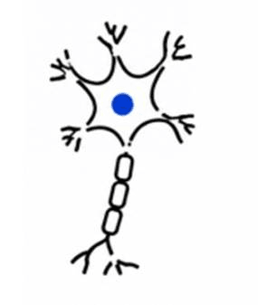 Neural Stem Cell (NSC)-derived Neurons
|
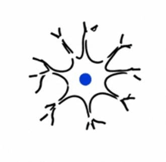 Neural Stem Cell (NSC)-derived Astrocytes
|
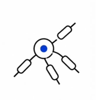 Neural Stem Cell (NSC)-derived Oligodendrocytes
|
Neural Stem Cell Growth Factors
| Growth Factor | English Name | Major Target / Signaling Pathway | Major Function | Main Applications |
|---|---|---|---|---|
| Epidermal Growth Factor | Epidermal Growth Factor (EGF) | EGFR → RAS/MAPK, PI3K/AKT signaling pathways | Promotes NSC/neural progenitor cell proliferation, maintains an undifferentiated state, and regulates glial lineage development | In vitro NSC expansion; neural progenitor maintenance; neural development studies |
| Fibroblast Growth Factor 2 | Fibroblast Growth Factor 2 (FGF2, bFGF) | FGFR1/2 → MAPK/ERK, PI3K/AKT, STAT pathways | Promotes NSC self-renewal and proliferation, maintains neural progenitor characteristics, and regulates adult neurogenesis | Core supplement for NSC expansion; iPSC/ESC-to-NSC induction systems; neural regeneration studies |
| Fibroblast Growth Factor 8 | Fibroblast Growth Factor 8 (FGF8) | FGFR → MAPK/ERK pathway | Regulates anterior–posterior neural tube patterning, midbrain regionalization, and neuronal subtype specification | Dopaminergic neuron induction; brain regional differentiation models |
| Brain-Derived Neurotrophic Factor | Brain-Derived Neurotrophic Factor (BDNF) | TrkB receptor → PI3K/AKT, MAPK/ERK, PLCγ pathways | Promotes neuronal survival, maturation, synapse formation, and neural network establishment | NSC-to-mature neuron differentiation; neuronal functional maturation; neurological disease models |
| Nerve Growth Factor | Nerve Growth Factor (NGF) | TrkA/p75NTR → MAPK, PI3K/AKT pathways | Promotes survival and axonal growth of sensory and sympathetic neurons | Neuronal culture; nerve injury repair studies |
| Insulin-like Growth Factor 1 | Insulin-like Growth Factor 1 (IGF-1) | IGF1R → PI3K/AKT, RAS/MAPK pathways | Promotes NSC survival, proliferation, and neurogenesis; enhances neuroprotective effects | Adult neurogenesis studies; neuroprotection and regeneration research |
| Vascular Endothelial Growth Factor | Vascular Endothelial Growth Factor (VEGF) | VEGFR2 → PI3K/AKT, MAPK pathways | Promotes NSC proliferation and neurogenesis; regulates the neurovascular microenvironment | Neural regeneration; brain injury repair; neurovascular coupling studies |
| Platelet-Derived Growth Factor-AA | Platelet-Derived Growth Factor-AA (PDGF-AA) | PDGFRα → PI3K/AKT, MAPK pathways | Promotes neural progenitor proliferation and oligodendrocyte lineage development | Oligodendrocyte induction; myelination studies |
| Sonic Hedgehog | Sonic Hedgehog (SHH) | PTCH1/SMO → GLI transcriptional pathway | Maintains NSC proliferation, regulates ventral neural tube patterning, and controls neuronal fate specification | Motor neuron induction; brain regionalization models; neural development studies |
| Wnt Proteins | Wnt Family Proteins (e.g., Wnt3a) | Frizzled/LRP5/6 → β-catenin pathway | Regulates NSC self-renewal, proliferation, neuronal differentiation, and neural pattern formation | Neural induction; neuronal subtype differentiation; brain organoid construction |
| Bone Morphogenetic Proteins | Bone Morphogenetic Proteins (BMP2/4/7) | BMPR → SMAD1/5/8 pathway | Regulates NSC differentiation fate, promotes astrocyte formation, and inhibits certain neuronal differentiation pathways | Glial cell induction; neural lineage fate studies |
| Noggin (BMP Inhibitor) | Noggin | BMP ligand inhibition → ↓SMAD signaling | Inhibits BMP-induced differentiation, promotes neural lineage maintenance and neural induction | hPSC-to-NSC induction (Dual SMAD inhibition); neural progenitor maintenance |
| Transforming Growth Factor β | Transforming Growth Factor β (TGF-β) | TGFβR → SMAD2/3 pathway | Regulates NSC quiescence, proliferation, and glial differentiation | Neural microenvironment studies; NSC fate regulation |
| Notch Ligands | Notch Ligands (DLL1/JAG1) | Notch receptor → NICD → Hes gene pathway | Maintains NSC undifferentiated state and prevents premature neuronal differentiation | Long-term NSC culture; neural development regulation studies |
Neural Stem Cell Markers
| Category | Marker | Significance |
|---|---|---|
| Core NSC marker | Nestin | Marker of neural progenitor cells |
| Core NSC marker | SOX2 | Maintains neural stem cell self-renewal capacity |
| NSC marker | PAX6 | Regulates neural lineage specification |
| Early neural progenitor marker | Musashi-1 | Maintains neural progenitor cell properties |
| Proliferation marker | Ki67 | Indicates active cell proliferation status |
| Neuronal differentiation marker | βIII-tubulin (TUJ1) | Marker of immature neurons |
| Mature neuronal marker | MAP2 | Marker of mature neurons |
| Astrocyte marker | GFAP | Marker of astrocyte lineage |
| Oligodendrocyte lineage marker | OLIG2 | Marker of oligodendrocyte lineage commitment |
Classical Identification Panels for Neural Stem Cells
| Cell Population | Typical Marker Combination | Characteristics |
|---|---|---|
| Undifferentiated Neural Stem Cells (NSCs) | Nestin⁺ / SOX2⁺ / PAX6⁺ / Musashi-1⁺ | Represents the NSC state with self-renewal capacity and multipotent differentiation potential. Nestin reflects neural progenitor characteristics, SOX2 maintains stemness and an undifferentiated state, PAX6 regulates neural lineage establishment, and Musashi-1 controls NSC proliferation and Notch-related signaling. Commonly used for quality assessment of hPSC-derived NSCs. |
| Neuroectoderm / Early Neural Progenitors | SOX1⁺ / PAX6⁺ / Nestin⁺ | Represents the early neural commitment stage from pluripotent stem cells toward the neural lineage, indicating the transition from pluripotency to neural fate. Commonly used to evaluate early-stage ESC/iPSC neural induction. |
| Radial Glial-like NSCs | SOX2⁺ / Nestin⁺ / GFAP⁺ / BLBP (FABP7)⁺ | Mimics embryonic radial glial characteristics, serving as an important source of neurons and glial cells during neural development. GFAP expression in some NSC populations reflects radial glial-like features. |
| Proliferative Neural Progenitor Cells (NPCs) | Nestin⁺ / SOX2⁺ / Ki67⁺ / EGFR⁺ | Represents highly proliferative neural progenitor populations. Ki67 indicates active cell-cycle status, while EGFR expression is associated with activated NSC/NPC proliferation. |
| NSC-enriched Population | CD133 (Prominin-1)⁺ / Nestin⁺ / SOX2⁺ | CD133 can be used for enrichment and sorting of certain NSC subpopulations; however, not all NSCs express CD133. Therefore, identification requires combination with Nestin, SOX2, and functional validation assays. |
| Adult Neural Stem Cells (Adult NSCs) | GFAP⁺ / SOX2⁺ / Nestin⁺ / BLBP⁺ | Mainly located in adult neurogenic regions, including the subventricular zone (SVZ) and subgranular zone (SGZ) of the hippocampus. These cells maintain long-term self-renewal capacity and generate neurons and glial cells. |
| Immature Neuronal Progenitors | DCX⁺ / PSA-NCAM⁺ / Low Nestin expression | Represents an intermediate stage during NSC-to-neuron differentiation, corresponding to migrating neuroblasts and immature neurons. |
| Early Neurons | TUJ1 (βIII-Tubulin)⁺ / DCX⁺ | Indicates initiation of the neuronal differentiation program from NSCs, accompanied by neurite extension and neuronal structural development. Commonly used to evaluate neuronal differentiation efficiency. |
| Mature Neurons | MAP2⁺ / NeuN⁺ / Synapsin-1⁺ | Represents functionally mature neurons with dendritic formation, neuronal-specific protein expression, and synaptic development capacity. Used for validation of terminal neuronal differentiation from NSCs. |
| Astrocyte Lineage | GFAP⁺ / S100β⁺ / ALDH1L1⁺ | Represents glial lineage differentiation from NSCs. GFAP is a classical astrocyte marker and is also expressed in some adult NSC populations. |
| Oligodendrocyte Lineage | OLIG2⁺ / PDGFRα⁺ / O4⁺ / MBP⁺ | Represents differentiation toward the oligodendrocyte lineage. OLIG2 and PDGFRα indicate early oligodendrocyte progenitors, while MBP represents mature myelin-forming oligodendrocytes. |
4 Mesenchymal Stem Cells, MSCs
Mesenchymal Stem Cells (MSCs) are adult stem cells derived from the mesoderm that possess self-renewal capacity, multilineage differentiation potential, and immunomodulatory functions. MSCs are widely distributed in various tissues, including bone marrow, adipose tissue, umbilical cord, and dental pulp, and serve as important seed cells for regenerative medicine and cell therapy research.
According to the criteria established by the International Society for Cell & Gene Therapy (ISCT), classical MSCs exhibit an adherent growth phenotype and consistently express mesenchymal markers, including CD73, CD90, and CD105, while lacking hematopoietic-associated markers such as CD45, CD34, CD14/CD11b, CD19, and HLA-DR. In addition, MSCs possess the ability to differentiate into osteoblasts, chondrocytes, and adipocytes, which represents a key functional criterion for evaluating their multipotency.
Beyond their traditional lineage differentiation capacity, MSCs exhibit strong paracrine regulatory and immunomodulatory functions. Through the secretion of cytokines, growth factors, extracellular vesicles, and other bioactive molecules, MSCs participate in inflammatory regulation, tissue repair, angiogenesis, and microenvironment remodeling. Therefore, MSCs and their derived products have been widely applied in tissue engineering, disease modeling, drug screening, immune regulation studies, and regenerative medicine research. Currently, MSC-based research platforms encompass multiple tissue sources, including bone marrow-derived MSCs (BM-MSCs), adipose-derived MSCs (AD-MSCs), and umbilical cord-derived MSCs (UC-MSCs). By optimizing culture systems, growth factor regulation, and lineage-specific induction strategies, MSC-derived functional cell populations with specialized characteristics can be further generated, providing reliable cellular resources for disease mechanism studies and the development of innovative therapeutic approaches.
Classical Characterization Markers
| Classification | Marker | English Name | Expression Characteristics | Main Significance |
|---|---|---|---|---|
| MSC Positive Markers | CD73 | Ecto-5′-nucleotidase | Highly expressed on the MSC surface | One of the core positive markers defined by ISCT; involved in purine metabolism and immunomodulation |
| MSC Positive Markers | CD90 | Thy-1 Cell Surface Antigen | Stably and highly expressed | Reflects MSC stemness and mesenchymal origin |
| MSC Positive Markers | CD105 | Endoglin | Highly expressed in undifferentiated MSCs | Associated with TGF-β signaling; maintains MSC proliferation and differentiation potential |
| MSC Positive Markers | CD44 | Hyaluronate Receptor | Broadly expressed | Involved in cell adhesion, migration, and tissue repair |
| MSC Positive Markers | CD29 | Integrin β1 | Highly expressed | Mediates extracellular matrix interaction and signal transduction |
| MSC Positive Markers | STRO-1 | Stromal Cell Marker-1 | Expressed in early MSC subpopulations | Commonly used for enrichment of MSCs with strong differentiation capacity |
| MSC Positive Markers | CD146 | Melanoma Cell Adhesion Molecule | Expressed in perivascular MSC-like populations | Associated with MSC migration, angiogenesis, and regenerative capacity |
| Stemness-Related Markers | Nestin | Neural Stem Cell Marker | Expressed in a subset of MSCs | Associated with MSC self-renewal and multipotent differentiation ability |
| Stemness-Related Markers | SSEA-4 | Stage-Specific Embryonic Antigen-4 | Expressed in some primitive MSC populations | Reflects a more primitive stem cell state |
| Negative Exclusion Markers | CD45 | Protein Tyrosine Phosphatase Receptor Type C | Not expressed | Excludes hematopoietic-derived cells |
| Negative Exclusion Markers | CD34 | Hematopoietic Progenitor Cell Antigen | Usually negative (may show weak expression in early MSCs) | Excludes hematopoietic progenitor cells |
| Negative Exclusion Markers | CD14/CD11b | Monocyte Marker | Not expressed | Excludes monocyte/macrophage lineage cells |
| Negative Exclusion Markers | CD19 | B Cell Marker | Not expressed | Excludes B cell-derived populations |
| Negative Exclusion Markers | HLA-DR | MHC Class II Molecule | Low or negative expression | Reflects the low immunogenicity of MSCs |
Growth Factors Related to Mesenchymal Stem Cell (MSC) Culture and Expansion
| Growth Factor | English Name | Major Target / Signaling Pathway | Effects on MSCs | Application Stage |
|---|---|---|---|---|
| FGF-2 | Fibroblast Growth Factor 2 | FGFR–MAPK/ERK pathway | Promotes MSC proliferation, improves expansion efficiency, and maintains an undifferentiated state | In vitro MSC expansion |
| PDGF-BB | Platelet-Derived Growth Factor-BB | PDGFR–PI3K/AKT pathway | Enhances MSC migration, proliferation, and vascular-related functions | Tissue repair research |
| EGF | Epidermal Growth Factor | EGFR–MAPK pathway | Enhances MSC proliferative capacity and improves cellular activity | MSC culture optimization |
| IGF-1 | Insulin-like Growth Factor 1 | IGF1R–PI3K/AKT pathway | Promotes cell survival, anti-apoptotic effects, and proliferation | MSC maintenance culture |
| TGF-β1 | Transforming Growth Factor Beta 1 | SMAD pathway | Regulates MSC fate determination and promotes chondrogenic differentiation | Differentiation induction |
| BMP-2 | Bone Morphogenetic Protein 2 | BMP–SMAD pathway | Strongly induces MSC differentiation into osteoblasts | Osteogenic induction |
| BMP-4 | Bone Morphogenetic Protein 4 | BMP pathway | Promotes osteogenic and chondrogenic lineage differentiation | Tissue engineering |
| VEGF | Vascular Endothelial Growth Factor | VEGFR pathway | Enhances MSC angiogenic functions | Vascular repair research |
| HGF | Hepatocyte Growth Factor | c-Met pathway | Enhances MSC migration, anti-inflammatory activity, and tissue repair capacity | Regenerative medicine |
| Wnt3a | Wingless-related Integration Site 3a | Wnt/β-catenin pathway | Regulates MSC self-renewal and lineage specification | Stemness maintenance |
| IL-6 | Interleukin-6 | JAK/STAT3 pathway | Regulates MSC immunomodulatory functions and inflammatory responses | Immunoregulation research |
Classical Identification Panel
| Category | Markers |
|---|---|
| Positive MSC markers | CD73, CD90, CD105 |
| Negative exclusion markers | CD34, CD45, CD14, CD19, HLA-DR |
| Stemness-related markers | STRO-1, Nestin, SSEA-4 |
| Differentiation validation markers | RUNX2 (osteogenic differentiation), PPARγ (adipogenic differentiation), SOX9 (chondrogenic differentiation) |
5 Cancer Stem Cells, CSCs
Cancer Stem Cells (CSCs) are a distinct subpopulation of cells present within tumor tissues that possess stem cell-like properties, including self-renewal capacity, multilineage differentiation potential, and the ability to sustain long-term tumor growth. The classical cancer stem cell theory proposes that tumors are not composed of a homogeneous population of identical cells, but rather consist of functionally diverse cellular subsets. Among these, CSCs serve as critical drivers of tumor initiation, progression, metastasis, and recurrence by generating highly heterogeneous tumor cell populations. Since the first identification of leukemia stem cells, CSC populations with tumor-initiating capacity have been subsequently identified in various solid tumors, including breast cancer, glioblastoma, colorectal cancer, hepatocellular carcinoma, pancreatic cancer, and lung cancer, and have been shown to be closely associated with therapeutic resistance.
Recent studies have demonstrated that CSC maintenance relies on complex molecular regulatory networks involving classical stemness-associated signaling pathways, including Wnt/β-catenin, Notch, Hedgehog, PI3K/AKT/mTOR, JAK/STAT, and TGF-β pathways. Meanwhile, components of the tumor microenvironment, such as hypoxia, inflammatory cytokines, cancer-associated fibroblasts (CAFs), immune cells, and extracellular matrix (ECM), can promote the acquisition of stem-like characteristics in tumor cells by regulating epigenetic states and cellular plasticity. Therefore, CSC research has gradually evolved from the traditional "rare specialized cell population model" toward a "dynamic tumor cell state transition model," emphasizing the ability of tumor cells to acquire and maintain stemness under different environmental stresses.
Cancer stem cells exhibit strong drug resistance and invasive potential, representing a major cause of residual disease, tumor recurrence, and distant metastasis after treatment. Targeting CSC-specific surface markers, such as CD133, CD44, EpCAM, ALDH1, and LGR5, as well as key regulatory pathways and CSC niches, has become an important strategy for precision oncology and the development of novel anticancer therapeutics. CSC culture systems derived from patient tumor samples, tumor organoid models, drug screening platforms, and functional validation systems provide powerful technological platforms for elucidating tumor initiation mechanisms, evaluating drug sensitivity, and developing personalized therapeutic strategies.