Homepage banner 1
Product Lines

Explore Our Portfolio

View all categories

Comprehensive Support

Learn More

Cell-Free Expression System Application

Cell-Free Protein Expression (CFPE) is a technology that enables the direct synthesis of proteins in an in vitro environment using cellular transcription-translation machinery extracted from living cells.

Explore Resources

CHO Cell Protein Expression Industrial Application

CHO cells are epithelial cells derived from the ovary of the Chinese hamster (Cricetulus griseus).

Explore Resources

HEK293 Cell Protein Expression Industrial Application

HEK293 cells and their derivatives have been widely used for the production of recombinant therapeutic proteins, vaccines, anticancer agents, and other clinically relevant drugs.

Explore Resources

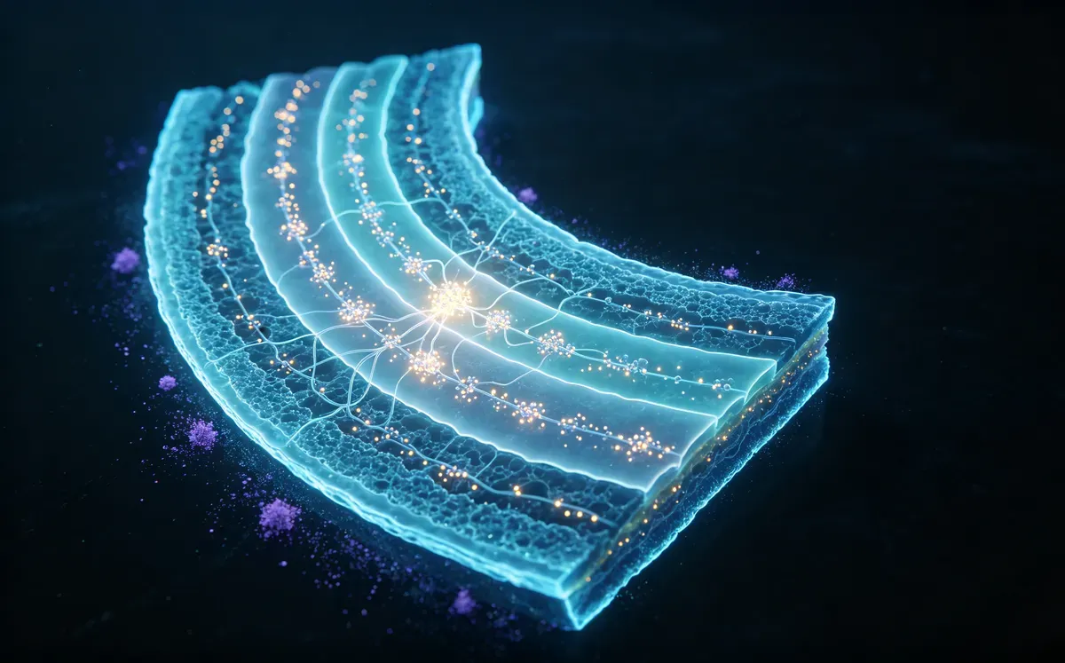
Antibody Solutions for Cortical Layers Marker Research

From Cortical Architecture to Neuronal Identity — Empowering Precision Neuroscience Research with Validated Layer-Specific Marker Antibodies

Explore Resources

Autophagy Signaling Research Solutions

Comprehensive autophagy research requires integrated analysis of autophagy initiation, autophagic flux, lysosomal function, selective organelle clearance, and metabolic signaling networks to understand cellular homeostasis, aging biology, and disease progression. We provide an integrated research toolkit — from autophagy core regulators to dynamic flux monitoring assays — enabling accurate investigation of autophagic activity and biological function. ## Key Target Highlights ![Autophagy Signaling Research Solutions-key.webp](https://cms.ucallm.com/uploads/Autophagy_Signaling_Research_Solutions_key_46fe5eadf6.webp) Key research trend: Modern autophagy research is increasingly moving beyond measuring single markers such as LC3-II accumulation toward comprehensive evaluation of autophagic flux, lysosomal competence, selective autophagy pathways, metabolic regulation, and organelle quality control. Emerging studies emphasize that autophagy functions as an adaptive signaling network integrating mTOR/AMPK nutrient sensing, mitochondrial homeostasis, immune regulation, and aging-associated stress responses, providing new opportunities for therapeutic intervention. ## Recommended Autophagy Marker Strategy ![Autophagy Signaling Research Solutions-reco1.webp](https://cms.ucallm.com/uploads/Autophagy_Signaling_Research_Solutions_reco1_48e064c06d.webp) ## Core Autophagy Validation Strategy ![Autophagy Signaling Research Solutions-core2.webp](https://cms.ucallm.com/uploads/Autophagy_Signaling_Research_Solutions_core2_b865d49232.webp) ## Competitive Technology Landscape ![Autophagy Signaling Research Solutions-compe.webp](https://cms.ucallm.com/uploads/Autophagy_Signaling_Research_Solutions_compe_fe47d223fe.webp) An integrated autophagy research workflow combining molecular validation, functional assays, advanced imaging, multi-omics profiling, and single-cell analysis to comprehensively characterize autophagy dynamics, mechanisms, and biological functions across multiple levels of biological complexity. ## Pathway Overview Autophagy is a conserved cellular recycling process that maintains homeostasis through degradation of damaged proteins and organelles. The ULK1 initiation complex, Beclin-1/VPS34 complex, ATG proteins, and lysosomal pathways coordinate autophagosome formation and cargo degradation. Autophagy regulates metabolism, stress adaptation, immunity, aging, and disease progression. ![Autophagy Signaling Research Solutions-path.webp](https://cms.ucallm.com/uploads/Autophagy_Signaling_Research_Solutions_path_1febe4225d.webp) Autophagy control by cellular signaling (PMID: 41086756) ## Recommended Experimental Validation Workflow ![Autophagy Signaling Research Solutions-reco2.webp](https://cms.ucallm.com/uploads/Autophagy_Signaling_Research_Solutions_reco2_466b248ef2.webp) ## Featured Research Application Examples ![Autophagy Signaling Research Solutions-fea.webp](https://cms.ucallm.com/uploads/Autophagy_Signaling_Research_Solutions_fea_7d3270c2e0.webp) ## Frequently Asked Questions Q1. Which biomarkers should I use to accurately assess autophagy? No single biomarker is sufficient. A comprehensive evaluation should combine LC3-I/II for autophagosome formation, SQSTM1/p62 for cargo degradation, Beclin-1 and ATG proteins for autophagy initiation, together with lysosomal markers such as LAMP1/2 to assess autophagosome maturation. .Q2. How can I distinguish autophagy induction from impaired autophagic degradation? Increased LC3-II alone does not necessarily indicate enhanced autophagy. Autophagic flux assays, combining lysosomal inhibitors (e.g., Bafilomycin A1 or Chloroquine) with LC3-II and p62 analysis, are considered the gold standard for differentiating increased autophagy from blocked lysosomal degradation. Q3. Which experimental approaches provide the most reliable assessment of autophagy? Robust autophagy studies integrate orthogonal methodologies, including Western blotting, immunofluorescence, autophagic flux assays, live-cell imaging, electron microscopy, and genetic perturbation. Combining complementary approaches minimizes experimental bias and strengthens biological conclusions. Q4. What are the most common pitfalls in autophagy research? Common pitfalls include interpreting LC3-II accumulation as increased autophagy without flux analysis, relying on a single biomarker, using insufficient experimental controls, and failing to validate antibody specificity. Multiple independent readouts and appropriate positive and negative controls are essential for accurate interpretation. Q5. How can autophagy be comprehensively characterized across biological systems? Modern autophagy research integrates protein validation, functional assays, high-resolution imaging, proteomics, metabolomics, and single-cell/spatial omics to reveal pathway activation, autophagic dynamics, functional remodeling, and cellular heterogeneity from molecular to tissue levels. ## Key References 1. Radulovic M., Yang C., Stenmark H. (2025).Lysosomal membrane homeostasis and its importance in physiology and disease.Nature Reviews Molecular Cell Biology. 27:71–87. 2. Nixon R.A., Rubinsztein D.C. (2024).Mechanisms of autophagy–lysosome dysfunction in neurodegenerative diseases.Nature Reviews Molecular Cell Biology. 25(12):926–946. 3. Settembre C., Perera R.M. (2024).Lysosomes as coordinators of cellular catabolism, metabolic signalling and organ physiology.Nature Reviews Molecular Cell Biology. 25:223–245. 4. Wang L., Klionsky D.J., Shen H.-M. (2023). The emerging mechanisms and functions of microautophagy. Nature Reviews Molecular Cell Biology. 24:186–203 5. Gross A.S., Ghillebert R., Schuetter M., et al. (2024). A metabolite sensor subunit of the Atg1/ULK complex regulates selective autophagy. Nature Cell Biology. 26. 6. Nakatogawa H. (2020). Mechanisms governing autophagosome biogenesis. Nature Reviews Molecular Cell Biology. 21:439–458. 7. Vargas J.N.S., Hamasaki M., Yoshimori T. (2022). The mechanisms and roles of selective autophagy in mammals. Nature Reviews Molecular Cell Biology. 24:167–185. 8. Kaur J., Debnath J. (2015). Autophagy at the crossroads of catabolism and anabolism. Nature Reviews Molecular Cell Biology. 16:461–472. 9. Klionsky D.J., Abdel-Aziz A.K., Abdelfatah S., et al. (2021). Guidelines for the use and interpretation of assays for monitoring autophagy (5th Edition). Autophagy. 10.

Explore Resources

Insulin Signaling Research Solutions

Insulin signaling regulates glucose metabolism, energy homeostasis, and cellular growth through pathways including PI3K–AKT–mTOR and MAPK signaling. Its dysregulation contributes to metabolic disorders, aging, cancer, and neurodegenerative diseases. We provide an integrated research platform — from insulin pathway targets to functional metabolic assays — supporting mechanistic studies of insulin resistance and metabolic regulation. ## Key Target Highlights ![Insulin Signaling Research Solutions-key.webp](https://cms.ucallm.com/uploads/Insulin_Signaling_Research_Solutions_key_ac4c329652.webp) Key research trend: Modern insulin signaling research is increasingly moving beyond the classical INSR–IRS–PI3K–AKT pathway toward integrated metabolic network analysis, combining nutrient sensing, inflammation, mitochondrial function, tissue-specific regulation, and aging-associated signaling to understand insulin resistance, metabolic disorders, and therapeutic opportunities. ## Recommended Insulin Signaling Marker Strategy ![Insulin Signaling Research Solutions-reco1.webp](https://cms.ucallm.com/uploads/Insulin_Signaling_Research_Solutions_reco1_35833d5f1e.webp) ## Core Insulin Signaling Validation Strategy ![Insulin Signaling Research Solutions-core.webp](https://cms.ucallm.com/uploads/Insulin_Signaling_Research_Solutions_core_446f0c38e2.webp) ## Competitive Technology Landscape ![Insulin Signaling Research Solutions-compe.webp](https://cms.ucallm.com/uploads/Insulin_Signaling_Research_Solutions_compe_a0644623c8.webp) An integrated insulin signaling research workflow combining pathway validation, functional assays, metabolic profiling, genetic analysis, disease modeling, and single-cell technologies to comprehensively define signaling mechanisms and metabolic regulation. ## Pathway Overview Insulin signaling is a central metabolic pathway controlling glucose uptake, lipid metabolism, protein synthesis, and cellular growth. Activation of the insulin receptor–IRS–PI3K–AKT–mTOR and MAPK pathways regulates metabolic homeostasis and cell survival. Dysregulation of insulin signaling contributes to insulin resistance, diabetes, aging-related disorders, cancer, and neurodegenerative diseases. ![Insulin Signaling Research Solutions-path.webp](https://cms.ucallm.com/uploads/Insulin_Signaling_Research_Solutions_path_476c5b9bda.webp) Overview of the main pathways of insulin signaling in regulating hepatic glucose and lipid metabolism (PMID: 38718757 ) ## Recommended Experimental Validation Workflow ![Insulin Signaling Research Solutions-reco2.webp](https://cms.ucallm.com/uploads/Insulin_Signaling_Research_Solutions_reco2_dac8c07883.webp) ## Featured Research Application Example ![Insulin Signaling Research Solutions-fea.webp](https://cms.ucallm.com/uploads/Insulin_Signaling_Research_Solutions_fea_a86a4fa182.webp) ## Frequently Asked Questions Q1.How can insulin signaling activity and metabolic function be accurately evaluated? A comprehensive evaluation integrates Western blotting or immunofluorescence for phosphorylation analysis with glucose uptake assays, glycogen synthesis assays, and Seahorse extracellular flux analysis to quantify glucose utilization, mitochondrial respiration, and glycolytic activity. Functional assays should complement molecular signaling measurements. Q2.What experimental approaches provide the most reliable analysis of insulin signaling? Robust studies combine orthogonal technologies, including phospho-protein analysis, genetic perturbation (CRISPR/RNAi), transcriptomics, metabolomics, live-cell imaging, and disease-relevant models such as organoids or genetically engineered animals. Integrating multiple approaches improves mechanistic interpretation and biological relevance. Q3.What are the most common pitfalls in insulin signaling research? Common challenges include suboptimal insulin stimulation conditions, inadequate serum starvation, relying on a single phospho-marker, insufficient biological controls, and overlooking pathway crosstalk with AMPK, mTOR, or MAPK signaling. Accurate interpretation requires optimized stimulation protocols, validated phospho-specific antibodies, and analysis of multiple signaling nodes. Q4.How can insulin signaling be comprehensively characterized in physiological and disease contexts? Modern insulin signaling research integrates pathway validation, metabolic phenotyping, genetic analysis, disease modeling, and single-cell/spatial omics to define signaling dynamics, metabolic regulation, tissue-specific responses, and cellular heterogeneity, providing systems-level insights into diabetes, obesity, cancer, and other metabolic disorders. ## Key References 1. Goul C, Peruzzo R, Zoncu R. (2023).The molecular basis of nutrient sensing and signalling by mTORC1 in metabolism regulation and disease. Nature Reviews Molecular Cell Biology. 24:857–875. 2. Choi E., Duan C., Bai X.-C. (2025). Regulation and function of insulin and insulin-like growth factor receptor signalling. Nature Reviews Molecular Cell Biology. 26:558–580. 3. Langer H.T., Rohm M., Sylow L. (2024). AMPK as a mediator of tissue preservation: time for a shift in dogma? Nature Reviews Endocrinology. 20. 4. Park J.B., Moon G.H., Cho A., et al. (2024). Neddylation of insulin receptor substrate acts as a bona fide regulator of insulin signaling and its implications for cancer cell migration. Cancer Gene Ther. 31:599–611. 5. Nature Reviews Endocrinology Editorial Collection. (2023–2025). Insulin signalling.

Explore Resources

New Arrivals

Learn More

U-Blot® PRG2 rabbit pAb

U-Blot® PRG2 rabbit pAb

View Product

U-Blot® MPP7 rabbit pAb

U-Blot® MPP7 rabbit pAb

View Product

U-Blot® Tuberin (phospho Thr1462) Polyclonal Antibody

U-Blot® Tuberin (phospho Thr1462) Polyclonal Antibody

View Product

U-Blot® Recombinant TNFa (VF0361) Fc-Fused Nanobody

U-Blot® Recombinant TNFa (VF0361) Fc-Fused Nanobody

View Product

U-Blot® Recombinant TNFa (VF0064) Fc-Fused Nanobody

U-Blot® Recombinant TNFa (VF0064) Fc-Fused Nanobody

View Product

U-Blot® Recombinant TIGIT (VF0242) Fc-Fused Nanobody

U-Blot® Recombinant TIGIT (VF0242) Fc-Fused Nanobody

View Product
Learn More

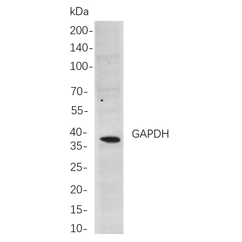
U-Blot® GAPDH Rabbit mAb

U-Blot® GAPDH Rabbit mAb

Learn More

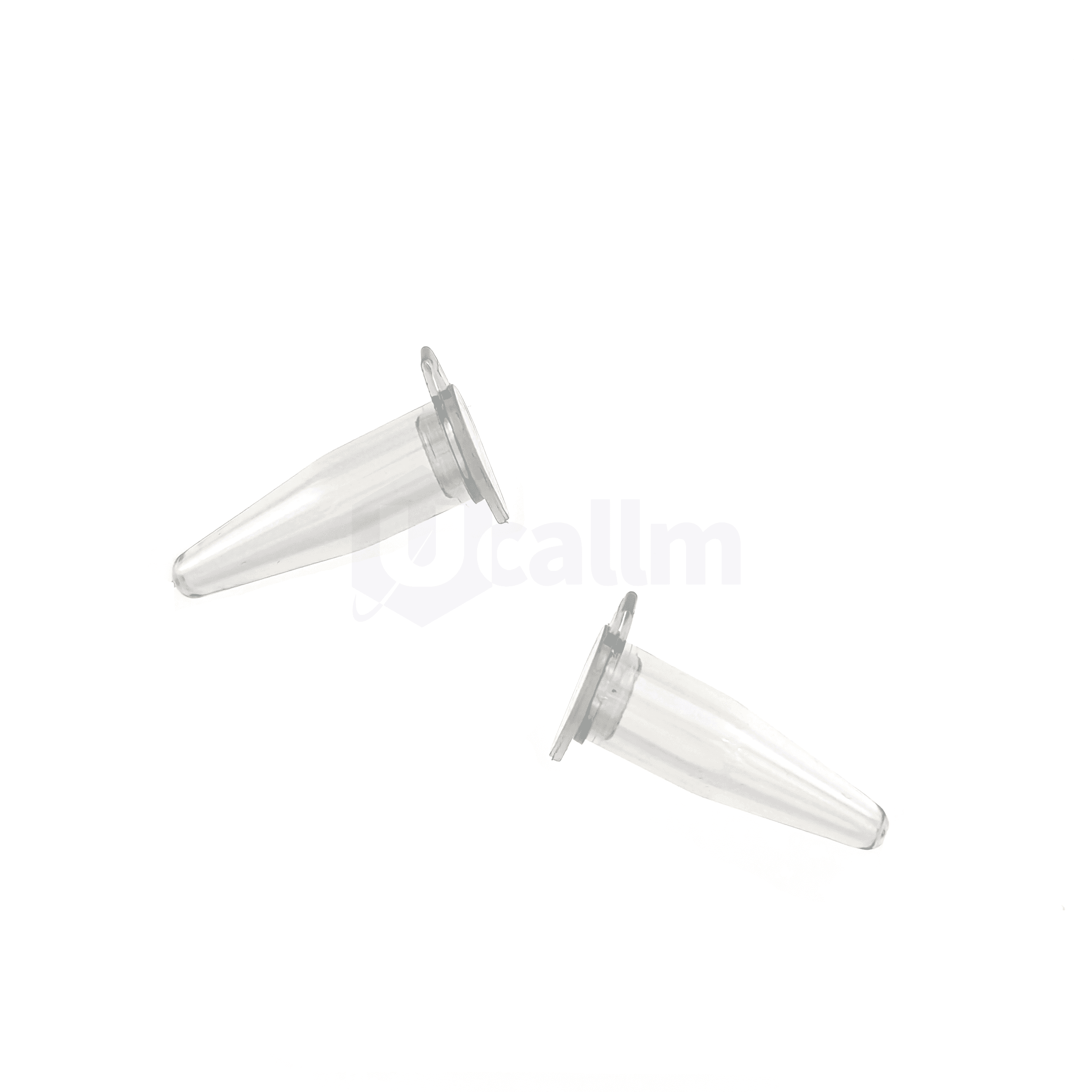
Thin-walled 0.2 mL Individual PCR Tube, with attached Optical Flat cap, Clear

PCR reaction vessels adopt ultra-pure medical-grade polypropylene used in the finest quality molding process to ensure the highest quality tubes, strips, caps, and microplates for PCR.

Learn More

8 Strip PCR Tube, plus strips of Optical flat caps, 0.2ml, clear PP wells, violet PC frame

PCR reaction vessels adopt ultra-pure medical-grade polypropylene used in the finest quality molding process to ensure the highest quality tubes, strips, caps, and microplates for PCR.

Learn More

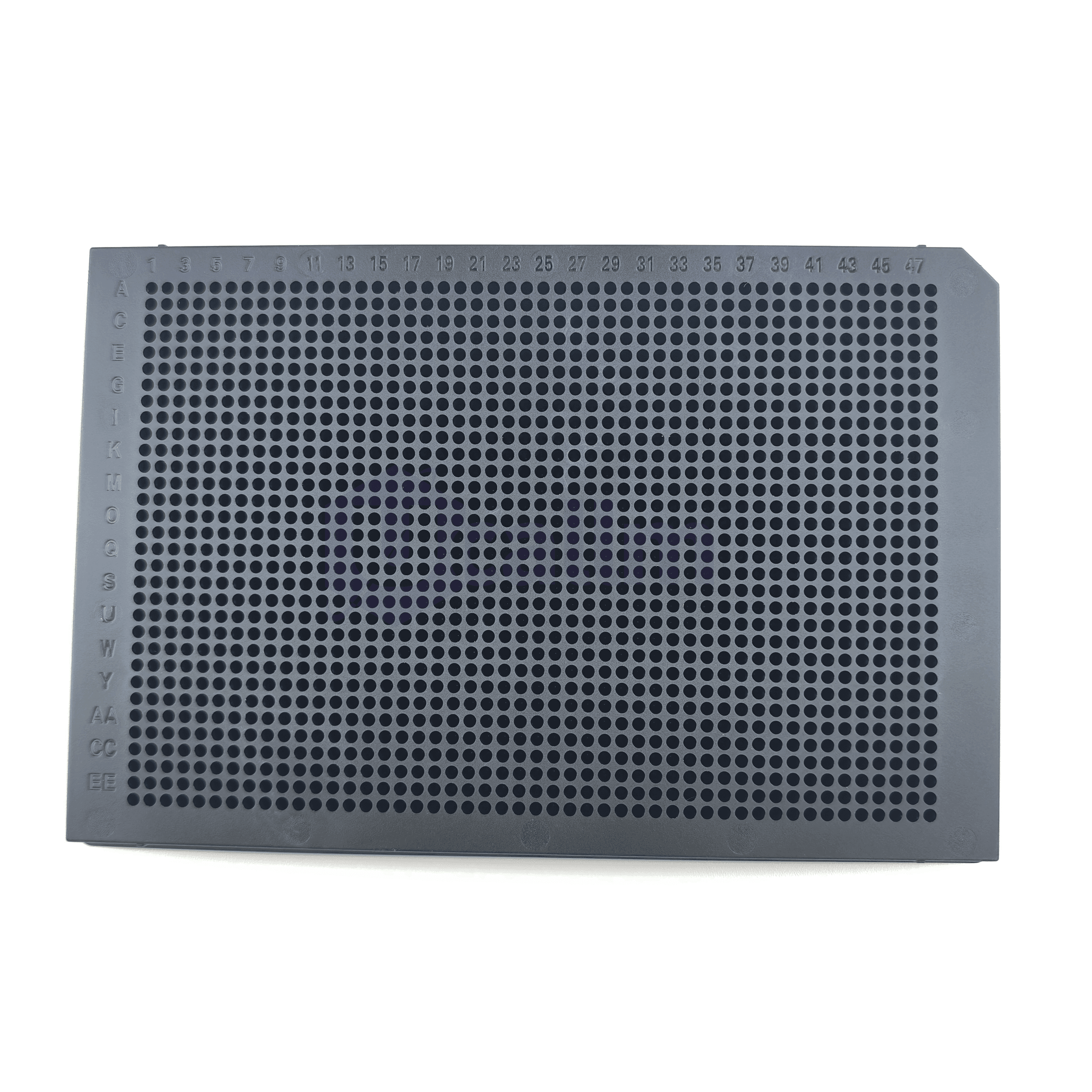
1536 Well PCR Plate, black

PCR reaction vessels adopt ultra-pure medical-grade polypropylene used in the finest quality molding process to ensure the highest quality tubes, strips, caps, and microplates for PCR.

Learn More

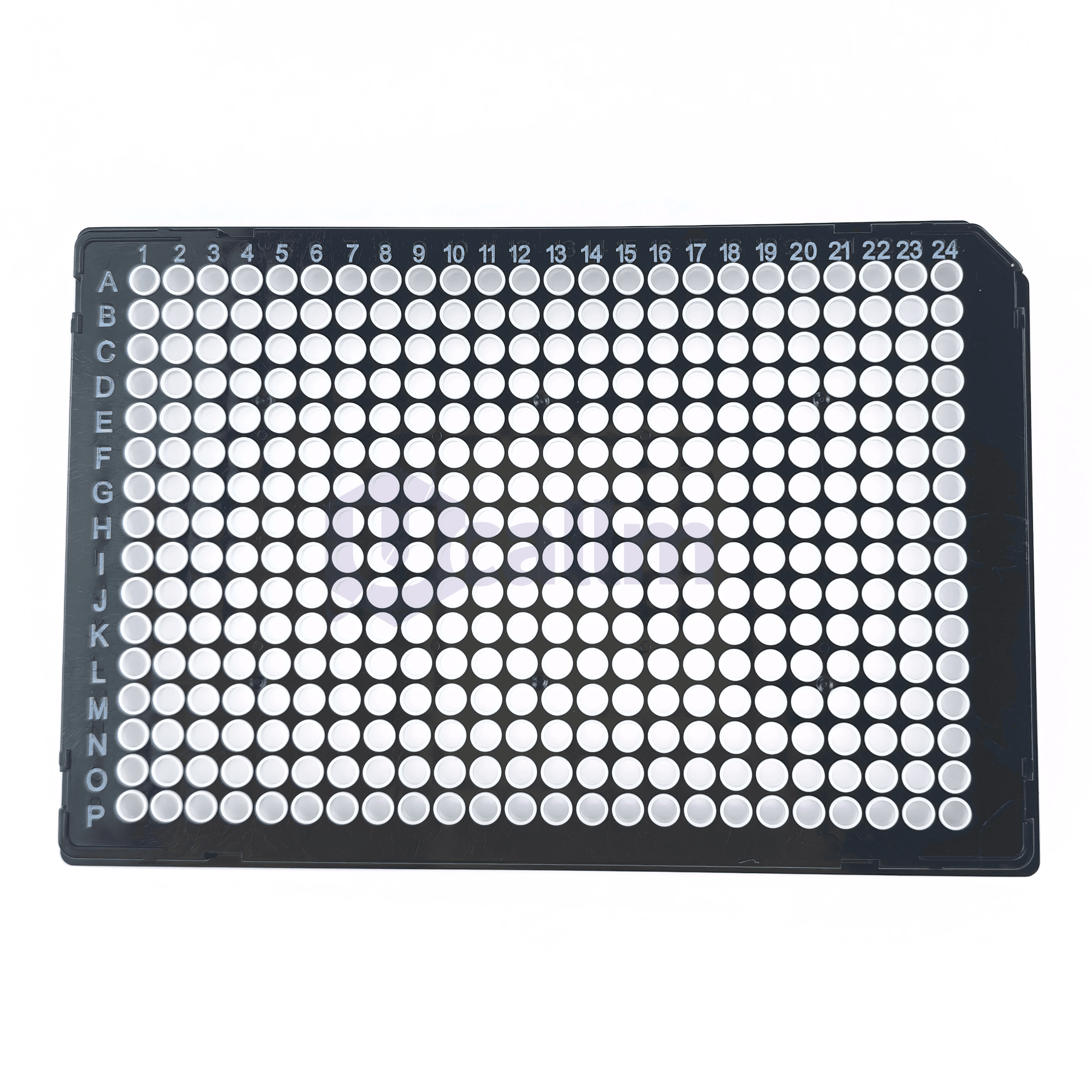
384 well Full skirt PCR plate, 40 µL, white PP wells, black PC frame, A24 Notch

PCR reaction vessels adopt ultra-pure medical-grade polypropylene used in the finest quality molding process to ensure the highest quality tubes, strips, caps, and microplates for PCR.

Learn More

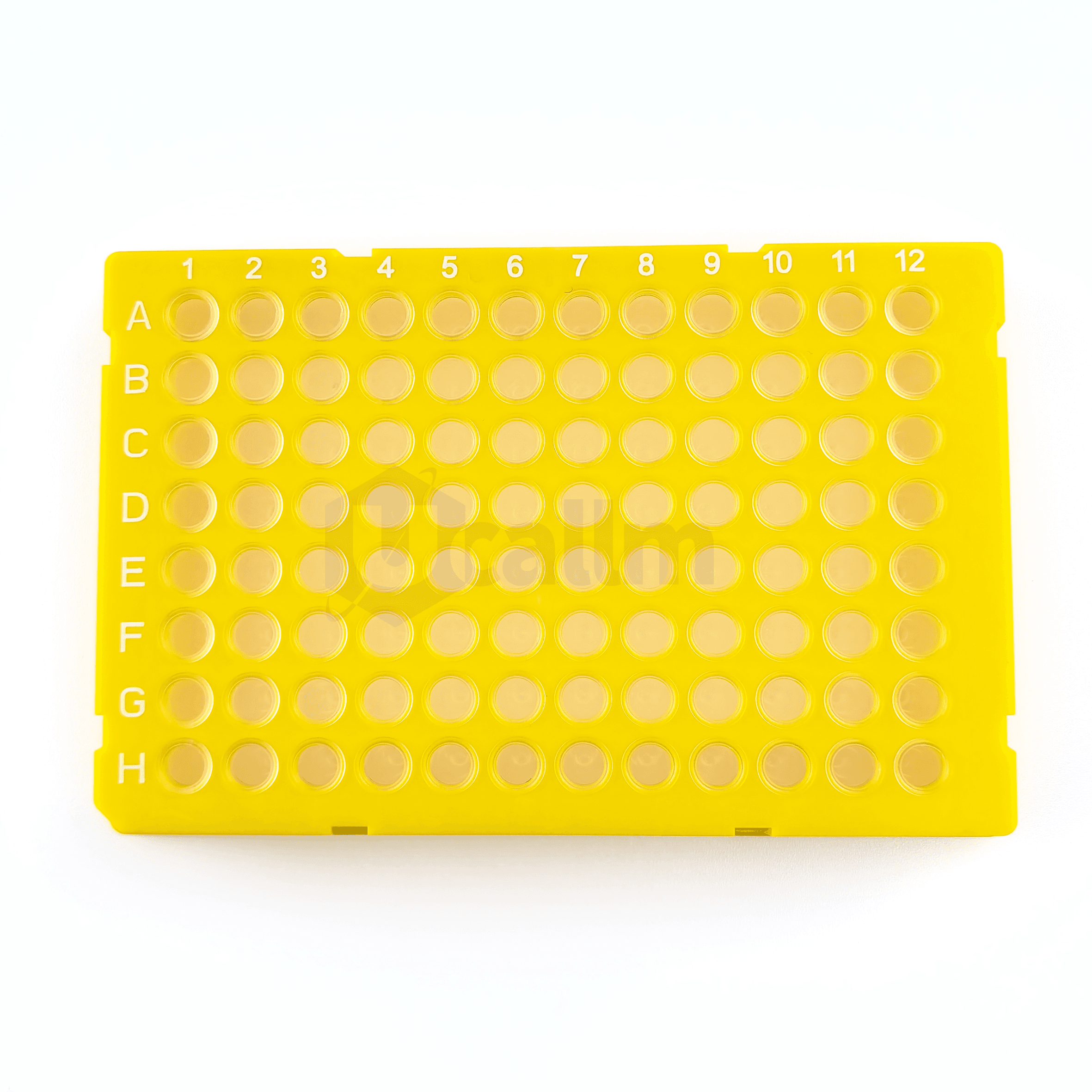
96 well Full skirt PCR plate, 0.1 mL, clear PP wells, yellow PC frame, H1 Notch

PCR reaction vessels adopt ultra-pure medical-grade polypropylene used in the finest quality molding process to ensure the highest quality tubes, strips, caps, and microplates for PCR.

Learn More

Search Products by Molecule Initial

Online Message