
info@ucallmlabs.com
From Neuroimmune Activation to Disease Mechanisms — Empowering Comprehensive Neuroinflammation Research with Validated Antibody Solutions
Neuroinflammation is a complex and highly dynamic process involving coordinated interactions among microglia, astrocytes, oligodendrocytes, neurons, infiltrating immune cells, and inflammatory signaling networks within the central nervous system (CNS).Neuroinflammatory mechanisms are implicated across a broad spectrum of neurological disorders, including Alzheimer’s disease (AD), Parkinson’s disease (PD), amyotrophic lateral sclerosis (ALS), multiple sclerosis (MS), stroke, traumatic brain injury (TBI), and brain tumors. Disease-associated changes encompass glial activation, innate and adaptive immune responses, inflammatory mediator release, altered CNS barrier function, and neuronal and myelin damage.
We enable comprehensive neuroinflammation research by supporting investigation across key biological frontiers, from microglial activation and astrocyte–microglia communication to neurodegeneration, synaptic dysfunction, and myelin repair. Validated research tools facilitate the study of immune-cell infiltration, neurovascular inflammation, and blood–brain barrier disruption, providing insights into complex CNS immune responses. Advanced multiplex immunofluorescence and spatial profiling technologies further reveal cellular interactions and tissue-level heterogeneity. Together, these approaches empower researchers to decode neuroimmune mechanisms, identify disease-associated biomarkers, and accelerate therapeutic discovery for neurological disorders.
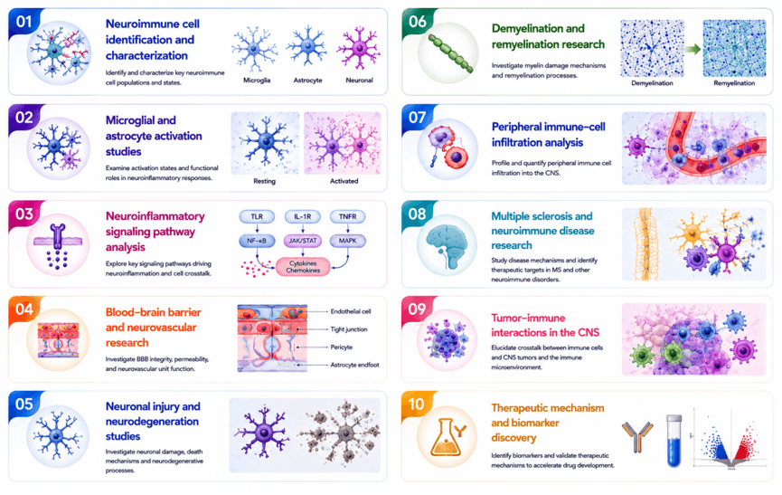
Neuroinflammation Biomarker Antibody Portfolio
—Comprehensive Solutions for Cellular Identification, Inflammatory Signaling, and Neurodegenerative Research
A multidimensional approach to neuroinflammation research requires simultaneous assessment of resident CNS cell populations, immune activation, inflammatory signaling, neural integrity, myelin status, and disease-associated pathways.
Research Area | Representative Biomarkers | Research Applications | Scientific Significance |
Microglial Identification & Activation | AIF1/Iba1, TMEM119, P2RY12, CD68, ITGAM/CD11b, HLA-DRA, P2RX4, P2RX7 | Microglial identification, activation-state characterization, neuroimmune signaling | Define resident microglial populations and investigate inflammatory phenotypes |
Astrocyte Identification & Reactivity | GFAP, S100B, GLUL, EZR, FABP7 | Astrocyte characterization and reactive gliosis studies | Evaluate astrocyte responses to CNS injury and inflammatory stimuli |
Neuronal Integrity & Injury | NEFM, NEFH, UCHL1, MAP2, GAP43 | Neuronal integrity, axonal injury, neurite remodeling | Monitor neuronal damage, axonal degeneration, and regenerative responses |
Oligodendrocyte & Myelin Biology | MBP, MOG, CNP, OLIG2, PLP1, SOX10 | Oligodendrocyte characterization, demyelination, remyelination | Assess myelin integrity and oligodendrocyte dysfunction |
Peripheral Immune Infiltration | CD3, CD4, CD8, CD20/MS4A1, ITGA4, GZMB, FOXP3 | T-cell, B-cell, and cytotoxic immune-cell profiling | Characterize adaptive immune responses within the CNS |
Inflammatory Signaling | IL-17A, IL-17RA, TLR2, TREM1, S100A8, S100A9, AGER/RAGE | Innate and adaptive inflammatory pathway studies | Investigate inflammatory mediator networks and immune activation |
Neurovascular & BBB Integrity | OCLN, CDH1/E-cadherin, EGFR, AGER | Blood–brain barrier and neurovascular research | Evaluate barrier integrity and inflammation-associated vascular alterations |
Neural Development & Cellular State | SOX2, SOX10, NES, RELN, NOTCH1, NR2E1/TLX | Neural progenitor biology and cellular-state analysis | Investigate neurogenesis, cellular plasticity, and disease-associated remodeling |
Neurodegenerative & Disease-Associated Pathways | TSPO, SORT1, EGR1, P2RX4/P2RX7, TLR2 | Neurodegeneration, mitochondrial stress, and inflammatory signaling | Connect cellular inflammatory responses with neurological disease mechanisms |
Key Neuroinflammation Biomarkers
——Molecular Functions, Research Applications, and Translational Value
Marker | Biological Function | Research Application | Translational Relevance |
AIF1/Iba1 | Actin-associated protein highly expressed in microglia and macrophages | Microglial identification and activation studies | Widely used marker for microglial responses |
TMEM119 | Microglia-associated membrane protein | Identification of resident microglia | Helps distinguish resident microglia from infiltrating myeloid populations |
P2RY12 | Purinergic receptor expressed by homeostatic microglia | Microglial state characterization | Sensitive indicator of microglial homeostatic alterations |
CD68 | Lysosomal glycoprotein associated with activated phagocytic cells | Phagocytic microglia/macrophage profiling | Supports assessment of inflammatory and phagocytic responses |
GFAP | Intermediate filament protein expressed by astrocytes | Astrocyte identification and reactive gliosis | Established marker of astrocyte activation and CNS injury |
MBP | Major structural component of CNS myelin | Demyelination and remyelination studies | Enables assessment of myelin integrity |
MOG | Myelin-associated oligodendrocyte protein | Oligodendrocyte and myelin research | Important in autoimmune demyelinating disease research |
NEFM | Neurofilament medium chain protein | Axonal integrity and neuronal injury | Supports evaluation of neuroaxonal damage |
MAP2 | Microtubule-associated neuronal cytoskeletal protein | Dendritic and neuronal integrity studies | Sensitive marker of neuronal structural alterations |
CD3 | Component of the T-cell receptor complex | Pan-T-cell identification | Enables characterization of CNS T-cell infiltration |
CD8 | Cytotoxic T-cell-associated surface protein | Cytotoxic immune-cell profiling | Supports analysis of adaptive immune responses |
CD20/MS4A1 | B-cell surface protein | B-cell identification and infiltration studies | Important for investigation of B-cell-mediated neuroinflammation |
FOXP3 | Transcription factor associated with regulatory T cells | Treg characterization | Enables investigation of immune suppression and regulation |
IL-17A | Pro-inflammatory cytokine produced by activated immune cells | Th17-associated inflammatory pathway studies | Relevant to autoimmune neuroinflammation, particularly MS |
TLR2 | Innate immune pattern-recognition receptor | Microglial and innate immune signaling | Investigates pathogen- and damage-associated inflammatory responses |
S100A8/S100A9 | Inflammatory calcium-binding proteins | Myeloid activation and inflammatory signaling | Associated with inflammatory immune-cell responses |
AGER/RAGE | Receptor mediating responses to advanced glycation end products and inflammatory ligands | Neuroinflammation and vascular signaling | Links inflammatory signaling with neurovascular and neurodegenerative processes |
TSPO | Mitochondrial membrane protein associated with activated glial responses | Neuroinflammation and neurodegeneration research | Widely investigated as a marker of glial activation |
Featured Neuroinflammation Research Panels
—Comprehensive Biomarker Panels for Cellular Profiling and Mechanistic Discovery
n Microglial Activation Panel—Defining Microglial Identity and Inflammatory State
Key Markers
TMEM119 | P2RY12 | AIF1/Iba1 | CD68 | HLA-DRA | P2RX7 |
n Astrocyte Reactivity Panel—Characterizing Astrocyte Responses to CNS Injury and Inflammation
Key Markers
GFAP | S100B | GLUL | FABP7 | EZR |
n Neurodegeneration & Neuronal Injury Panel—Linking Neuroinflammation to Neuronal and Axonal Damage
Key Markers
NEFM | NEFH | MAP2 | GAP43 | GFAP | UCHL1 |
n Demyelination & Oligodendrocyte Panel—Assessing Myelin Integrity and Neuroimmune-Mediated Damage
Key Markers
MBP | MOG | PLP1 | CNP | OLIG2 | SOX10 |
n Neuroimmune Infiltration Panel—Characterizing Adaptive Immune Responses in the CNS
Key Markers
CD3 | CD4 | CD8 | CD20 | FOXP3 | GZMB |
Neuroinflammation Research
From Cellular Identification to Mechanistic and Translational Discovery
We provide comprehensive research solutions for investigating neuroinflammation from cellular characterization to mechanistic discovery and biomarker translation. By combining validated markers for glial identity, immune infiltration, inflammatory signaling, neuronal injury, and myelin integrity, researchers can systematically profile neuroimmune interactions within the CNS. Advanced multiplex imaging and spatial profiling approaches further enable visualization of complex cellular networks and disease-associated pathways. These integrated tools support the study of neurodegenerative diseases, autoimmune disorders, and CNS injury, accelerating the identification of novel biomarkers and therapeutic targets for precision neuroinflammation research.
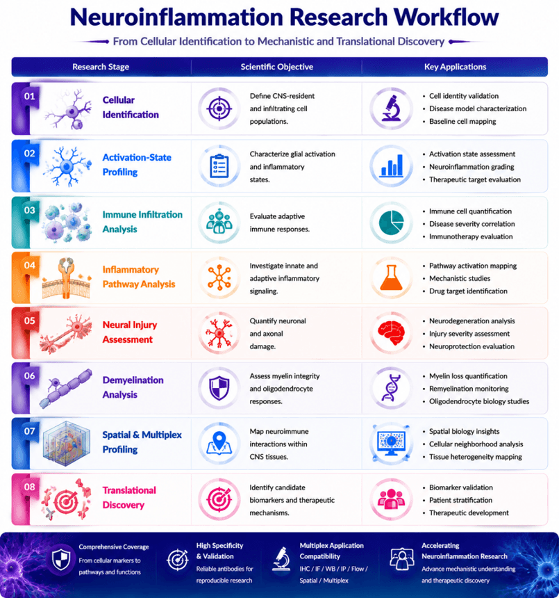
Why Researchers Choose Our Neuroinflammation Antibody Solutions
Validated Targets. Comprehensive Panels. Reliable Neuroimmune Research.
Our antibody portfolio supports the complete neuroinflammation research workflow—from cellular identification and activation-state profiling to pathway interrogation, tissue pathology, and translational biomarker discovery.
Research Challenge
Our Solution
How can I identify activated microglia?
Comprehensive microglial markers including AIF1/Iba1, TMEM119, P2RY12, CD68, and HLA-DRA
How can I distinguish CNS cell populations?
Integrated markers for microglia, astrocytes, neurons, oligodendrocytes, and infiltrating immune cells
How can I evaluate neuroinflammatory signaling?
Targeted antibodies against TLR2, P2RX4/P2RX7, IL-17A/IL-17RA, TREM1, and AGER/RAGE
How can I assess neuronal damage?
Neuronal and axonal markers including NEFM, NEFH, MAP2, UCHL1, and GAP43
How can I investigate demyelination?
Myelin and oligodendrocyte markers including MBP, MOG, PLP1, CNP, OLIG2, and SOX10
How can I characterize CNS immune infiltration?
Immune-cell markers including CD3, CD4, CD8, CD20, FOXP3, ITGA4, and GZMB
How can I resolve complex neuroimmune interactions?
Multiplex-compatible marker combinations for spatial characterization of cellular and molecular phenotypes
How can I improve experimental reproducibility?
Validated antibody solutions optimized for IHC, ICC/IF, WB, flow cytometry, and multiplex applications
Key References
1. Nixon R.A., Rubinsztein D.C. (2024)Mechanisms of autophagy–lysosome dysfunction in neurodegenerative diseases. Nature Reviews Molecular Cell Biology, 25:926–946.
2. Woodburn S.C., Bollinger J.L., Wohleb E.S. (2021)The semantics of microglia activation: neuroinflammation, homeostasis, and stress. Journal of Neuroinflammation, 18:258.
3. Leng F., Edison P. (2021)Neuroinflammation and microglial activation in Alzheimer disease: where do we go from here? Nature Reviews Neurology, 17:157–172.
4. Zong S. et al. (2026)Microglia and neuroinflammation: function, heterogeneity, and crosstalk Cellular & Molecular Immunology, 23:787–805.
5. Ajoolabady A. et al. (2025) Dual role of microglia in neuroinflammation and neurodegenerative diseases. Neurobiology of Disease, 216:107133.