
info@ucallmlabs.com
1.Overview
Ebola virus (EBOV) is the prototype species of the genus Orthoebolavirus within the family Filoviridae and is the causative agent of Ebola virus disease (EVD). It is an enveloped, negative‑sense, single‑stranded, non‑segmented RNA virus with filamentous or fibrous virions, approximately 80 nm in diameter and 650–1,400 nm in length.
Ebola virus is one of the most pathogenic viruses known to infect humans, with case fatality rates ranging from 25% to 90%. First identified in 1976 in Zaire (now Democratic Republic of the Congo) and Sudan, the virus has caused multiple major outbreaks in Africa. The 2013–2016 West African epidemic was the largest in history, with 28,646 cases and 11,323 deaths documented. Between 2021 and 2025, consecutive filoviral outbreaks have occurred.
The genus Orthoebolavirus comprises six distinct virus species, among which Zaire ebolavirus (EBOV), Sudan virus (SUDV), and Bundibugyo virus (BDBV) have caused significant outbreaks in humans. Fruit bats are considered the natural reservoir of Ebola virus, while non‑human primates and other animal species may also play a role in transmission.
2.Background Information
2.1 What is Ebola Virus?
Ebola virus is an enveloped, negative‑sense, single‑stranded RNA virus belonging to the family Filoviridae and genus Orthoebolavirus. The virion is filamentous or fibrous, approximately 80 nm in diameter and 650–1,400 nm in length. The viral envelope is derived from the host cell membrane and contains trimeric glycoprotein (GP) spikes responsible for receptor recognition and membrane fusion. The inner side of the envelope is lined by the matrix protein VP40.
The Ebola virus genome is a negative‑sense single‑stranded RNA of approximately 18.9 kb. The genome encodes seven genes in the order 3′ to 5′, each flanked by 3′ and 5′ untranslated regions.
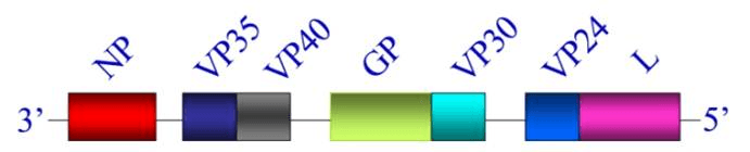
EBOV genome. The genes are depicted as boxes: nucleoprotein (NP), viral protein (VP) 35, matrix protein VP40, glycoprotein (GP), VP30, VP24, and polymerase protein (L). (PMID: 28903457)
2.2 Ebola Virus Genome & Classification
Ebola virus belongs to the genus Orthoebolavirus in the family Filoviridae. Its genome is a negative‑sense single‑stranded RNA of approximately 18.9 kb, encoding seven genes. The gene order is: 3′‑NP‑VP35‑VP40‑GP‑VP30‑VP24‑L‑5′. Each gene is flanked by 3′ and 5′ untranslated regions containing start and stop signals.
The family Filoviridae currently encompasses six distinct virus species. Those that have caused significant outbreaks in humans include:
Zaire ebolavirus (EBOV) : highest case fatality rate, up to 88%
Sudan virus (SUDV) : case fatality rate approximately 50%
Bundibugyo virus (BDBV) : case fatality rate approximately 25–35%
2.3 What Are Ebola Virus Structural Proteins?
Ebola virus encodes seven structural proteins with the following major functions:
| Protein | Full name | Main Function |
| NP | Nucleoprotein | Binds viral RNA, forms the nucleocapsid core structure |
| VP35 | Viral protein 35 | Polymerase cofactor, involved in transcription/replication, antagonizes host interferon responses |
| VP40 | Viral protein 40 | Matrix protein, mediates viral assembly and budding |
| GP | Glycoprotein | Surface spike protein, mediates receptor binding and membrane fusion |
| VP30 | Viral protein 30 | Transcriptional activator |
| VP24 | Viral protein 24 | Nucleocapsid component, regulates nucleocapsid assembly and function |
| L | Large protein | RNA‑dependent RNA polymerase (RdRp) |
2.3.1 Nucleocapsid Structure
Filamentous Ebola virions contain a helical nucleocapsid responsible for genome transcription, replication, and packaging into progeny virions. The nucleocapsid consists of a helical nucleoprotein (NP)–viral genomic RNA complex forming the core structure, to which VP24 and VP35 bind externally. Two NPs, each paired with a VP24 molecule, constitute a repeating unit.
A 2025 study using single‑particle cryo‑electron microscopy determined the nucleocapsid‑like structure within virus‑like particles at 4.6 Å resolution. The study demonstrated that the two VP24s in different orientations distinctively regulate nucleocapsid assembly, viral RNA synthesis, intracellular transport of the nucleocapsid, and infectious virion production.
2.3.2 Function of VP40
VP40 is the matrix protein of Ebola virus, forming a matrix layer beneath the viral surface that provides stability and shape. VP40 predominantly forms a dimer, binds to the host cell plasma membrane inner leaflet, and serves as a building block for matrix layer formation. VP40 also triggers inflammatory responses by activating the p65‑dependent canonical NF‑κB pathway, which may be linked to Ebola virus virulence.
2.4 Mechanism of Cell Entry and Replication
2.4.1 Viral Attachment and Receptor Recognition
Ebola virus infection begins with the binding of viral GP protein to multiple receptors on the host cell surface. GP exists as trimers on the viral envelope, responsible for recognizing host receptors and mediating membrane fusion.
2.4.2 Viral Entry
Ebola virus enters host cells primarily through macropinocytosis. After internalization, the virus is trafficked through endosomes to late endosomes/lysosomes. During this process, host proteases such as cathepsin B cleave GP, exposing the receptor‑binding site. The cleaved GP then binds to the host endosomal receptor Niemann‑Pick C1 (NPC1) , triggering membrane fusion and release of the viral nucleocapsid into the cytoplasm. NPC1 is an obligate host receptor required for Ebola virus entry.
2.4.3 Viral RNA Replication
After nucleocapsid release into the cytoplasm, the L protein (RdRp) carried by the virion, together with its cofactor VP35, initiates transcription and replication of the genome. Ebola virus replicates entirely within the host cell cytoplasm. Replication involves:
Transcription: negative‑sense RNA genome is transcribed into mRNAs, which are translated into viral proteins
Replication: positive‑sense RNA intermediates are synthesized using negative‑sense RNA as template
Progeny negative‑sense RNA genomes are synthesized using positive‑sense RNA as template
Newly synthesized NP protein immediately binds to progeny negative‑sense RNA to form ribonucleoprotein complexes (RNPs).
2.4.4 Viral Assembly and Release
Viral assembly begins with the interaction of VP40 with the inner leaflet of the plasma membrane. VP40 selectively interacts with phosphatidylserine (PS) and phosphatidylinositol‑4,5‑bisphosphate (PI(4,5)P2). VP40 connects the nucleocapsid to regions of the cell membrane enriched with GP. The virus acquires its envelope by budding from the host cell membrane, forming filamentous virions that are released.
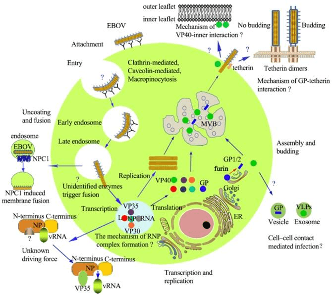
The life cycle of Ebola virus. (PMID: 28903457)
2.5 Symptoms and Treatment
2.5.1 Clinical Symptoms
The incubation period for Ebola virus disease is typically 2–21 days. Clinical presentation ranges from asymptomatic infection to severe hemorrhagic fever. Common symptoms include:
Acute onset of fever and severe headache
Muscle pain and weakness
Sore throat, cough
Vomiting, diarrhea, and abdominal pain
Rash
Unexplained hemorrhage (internal or external)
Multi‑organ dysfunction in advanced stages
Severe cases may involve severe hemorrhage, shock, acute respiratory distress syndrome, and multi‑organ failure. Ebola virus disease is one of the deadliest viral diseases known.
2.5.2 Antiviral Treatment
Treatment of Ebola virus disease is primarily supportive, including:
Intravenous fluid resuscitation: to maintain blood pressure and electrolyte balance
Oxygen therapy
Blood pressure stabilization
Management of complications: bleeding, renal failure, etc.
Two monoclonal antibody therapies are currently FDA‑approved for the treatment of Zaire Ebola virus infection:
REGN-EB3: a combination of three monoclonal antibodies (Atoltivimab, Maftivimab, Odesivimab)
mAb-114 (Ansuvimab) : a human monoclonal antibody derived from a 1995 outbreak survivor
Monoclonal antibodies have become the standard of care for Ebola virus disease. Additionally, Remdesivir has shown partial protection in non‑human primate models.
2.6 Targets for Intervention
2.6.1 Viral Targets
| Target | Protein | Biological Role |
| GP | Glycoprotein | Receptor binding & membrane fusion |
| L protein | RNA polymerase (L) | RNA transcription & replication |
| VP35 | Polymerase cofactor | Transcription/replication, interferon antagonism |
| VP40 | Matrix protein | Assembly & budding |
| VP24 | Nucleocapsid regulator | Nucleocapsid assembly & function |
| NP | Nucleoprotein | RNA binding & nucleocapsid formation |
2.6.2 Host Receptors & Entry Factors
| Host Target | Type | Role in Infection |
| NPC1 | Endosomal receptor | Obligate receptor for viral entry |
| Cathepsin B | Cysteine protease | GP proteolytic cleavage activation |
| Macropinocytosis | Endocytic pathway | Viral entry |
| CXCR4 | Chemokine receptor | Promotes viral entry |
A 2025 study revealed that CXCR4 plays a dual role in Ebola virus infection: promoting viral entry while also downregulating cytotoxic GP to promote viral fitness.
2.6.3 Host Signaling Pathways
| Pathway | Key Targets | Biological Role |
| NF‑κB pathway | p65, IKK | Inflammatory response, activated by VP40 |
| Interferon pathway | IFN‑α/β | Antiviral response, suppressed by VP35 |
| RIG‑I signaling | RIG‑I | Viral RNA sensing |
2.7 Vaccine Types and Development Progress
Two Ebola vaccines are currently licensed, both targeting Zaire ebolavirus:
2.7.1 Ervebo (rVSVΔG-ZEBOV-GP)
Ervebo is a single‑dose live‑attenuated vesicular stomatitis virus (VSV)‑vectored vaccine expressing the Zaire ebolavirus glycoprotein. The vaccine demonstrated near‑complete protection in ring vaccination campaigns in Guinea and the Democratic Republic of the Congo. Real‑world effectiveness is 84% (95% CI 70–92). Antibodies persist for up to two years post‑vaccination.
2.7.2 Zabdeno/Mvabea (Ad26.ZEBOV/MVA-BN-Filo)
A two‑dose heterologous prime‑boost regimen: first dose Ad26.ZEBOV (adenovirus‑vectored), second dose MVA‑BN‑Filo (modified vaccinia Ankara‑vectored).
2.7.3 Bivalent ChAdOx1-Vectored Vaccine
A Phase 1 clinical trial published in The Lancet Microbe in 2025 evaluated the safety and immunogenicity of a bivalent ChAdOx1‑vectored vaccine targeting both Ebola virus and Sudan virus. This was a first‑in‑human, open‑label, non‑randomised Phase 1 trial in healthy UK adults.
2.7.4 GamEvac-Combi (Heterologous rVSV and rAd5-Vectored Vaccine)
A 2025 randomized controlled multicenter trial in the Republic of Guinea and Russia evaluated GamEvac‑Combi. Results showed:
Marked GP‑specific IFN‑γ response at day 28
Neutralizing response at day 42 with seroconversion rate of 96.3% (GMT = 32.6)
GP‑specific IgG antibody levels peaked at day 42 (GMT = 9345) and persisted for at least one year (GMT = 650)
2.7.5 Sudan Ebola Virus Vaccine
In February 2025, Uganda's Ministry of Health, WHO and partners launched the first‑ever clinical efficacy trial for a vaccine against Ebola Sudan virus. This is the first trial to assess the clinical efficacy of a vaccine against Ebola Sudan virus disease. The first 2,160 doses of the vaccine candidate were pre‑positioned in Kampala as part of outbreak preparedness. The trial was launched just four days after outbreak confirmation, setting a speed record for emergency randomized vaccine trials.
2.7.6 mRNA Vaccines
A 2025 study reported that an mRNA vaccine encoding the Ebola virus glycoprotein induced high neutralizing antibody titers and provided strong protection against lethal infection in mouse models.
2.7.7 Vaccination in Children
A systematic review and meta-analysis showed that in children, seroconversion after the first dose of Ebola vaccine was 89%, rising to 96% after full vaccination.
2.8 Drugs
Currently, no small‑molecule antiviral drug is specifically approved for Ebola virus. Treatment relies primarily on supportive care and monoclonal antibody therapy. Based on their targets, they can be classified as follows:
2.8.1 Virus-Targeted Drugs
| Target | Drug | Mechanism |
| GP | Monoclonal antibodies (REGN‑EB3, mAb‑114) | Neutralization |
| Novel mAb 3A6 | Binds GP, protects highly viremic animals | |
L protein (RdRp) | Remdesivir | RNA synthesis inhibition |
| Galidesivir | RNA synthesis inhibition | |
| Favipiravir | RNA synthesis inhibition | |
| Obeldesivir | Oral RdRp inhibitor | |
| Entry | Novel tetrahydroisoquinoline entry inhibitor (Hu7) | Blocks viral entry |
| Diarylamine derivatives | Blocks viral entry | |
| Probenecid | Inhibits filovirus replication | |
| Toremifene, Clomiphene | GP inhibitors | |
| Cathepsin B | CA‑074 | Cathepsin B inhibition |
CA-074: A 2025 comprehensive in silico study evaluated six small‑molecule inhibitors. CA‑074 was identified as the most promising candidate, exhibiting strong binding affinity to cathepsin B (−40.87 kcal/mol), an endosomal cysteine protease crucial for Ebola virus entry, along with favorable ADMET properties and safety indicators. Molecular dynamics confirmed the stability of the CA‑074–cathepsin B complex over 300 ns. CA‑074 has the potential to be a leading candidate for the treatment of EBOV.
Novel entry inhibitors: A 2025 study reported a series of novel Ebola entry inhibitors based on the 1,2,3,4‑tetrahydroisoquinoline‑3‑carboxamide scaffold. Compound Hu7 demonstrated antiviral activity comparable to previous findings with markedly reduced toxicity.
2.8.2 Host-Targeted Drugs
| Target | Drug | Mechanism | Stage |
| Immune modulation | Monoclonal antibodies | Passive immunization | FDA‑approved |
| Supportive care | IV fluids, oxygen therapy | Supportive care | Standard of care |
2.9 Viral Persistence
2025 studies have demonstrated that Ebola virus can persist in survivors for months. Research on Sudan ebolavirus disease survivors found:
50% of survivors reported persistent multi‑systemic symptoms two years post‑infection
Viral RNA could be detected in semen and breast milk for up to 7 months
Long‑term sequelae include chronic musculoskeletal, sensory, and neurological symptoms
These findings suggest the possibility of viral latency and reactivation, with important implications for outbreak control and long‑term survivor health management.
2.10 Reverse Genetics Systems
Reverse genetics systems are important tools for Ebola virus research. Several advances were reported in 2025:
Development of a pentacistronic Ebola virus minigenome system
Recombinant measles virus‑vectored Ebola vaccine construction and reverse genetics system
Genome‑scale CRISPR knockout screens in Ebola virus replicon cell lines
3.Related Products & Services
Recombinant Antigens & Receptors
Antibodies
Detection Kits
Recombinant Protein Expression Services
Antibody Development Services
4.Resources
5.References
Feldmann, H., Geisbert, T. W. (2011). Ebola haemorrhagic fever. The Lancet, 377, 849–862.
Baseler, L., Chertow, D. S., Johnson, K. M., et al. (2017). The pathogenesis of Ebola virus disease. Annual Review of Pathology: Mechanisms of Disease, 12, 387–418.
Carette, J. E., Raaben, M., Wong, A. C., et al. (2011). Ebola virus entry requires the cholesterol transporter Niemann–Pick C1. Nature, 477, 340–343.
Chandran, K., Sullivan, N. J., Felbor, U., et al. (2005). Endosomal proteolysis of the Ebola virus glycoprotein is necessary for infection. Science, 308, 1643–1645.
Bharat, T. A. M., Noda, T., Riches, J. D., et al. (2012). Structural dissection of Ebola virus and its assembly. Nature, 477, 330–334.
Martin-Serrano, J., Zang, T., Bieniasz, P. D. (2001). HIV-1 and Ebola virus encode small peptide motifs that recruit Tsg101 to sites of particle assembly to facilitate egress. Nature Medicine, 7, 1313–1319.
Hoenen, T., Groseth, A., Feldmann, H. (2019). Therapeutic strategies to target the Ebola virus life cycle. Nature Reviews Microbiology, 17, 593–606.
Mulangu, S., Dodd, L. E., Davey, R. T., et al. (2019). A randomized, controlled trial of Ebola virus disease therapeutics. New England Journal of Medicine, 381, 2293–2303.
Warren, T. K., Jordan, R., Lo, M. K., et al. (2016). Therapeutic efficacy of the small molecule GS-5734 against Ebola virus in rhesus monkeys. Nature, 531, 381–385.
Warren, T. K., Jordan, R., Lo, M. K., et al. (2016). Therapeutic efficacy of the small molecule GS-5734 against Ebola virus in rhesus monkeys. Nature, 531, 381–385.
Regules, J. A., Beigel, J. H., Paolino, K. M., et al. (2017). A recombinant vesicular stomatitis virus Ebola vaccine. New England Journal of Medicine, 376, 330–341.
Kennedy, S. B., Bolay, F., Kieh, M., et al. (2017). Phase 2 placebo-controlled trial of two vaccines to prevent Ebola in Liberia. New England Journal of Medicine, 377, 1438–1447.
Halperin, S. A., Das, R., Mao, L., et al. (2023). Randomized trial of vaccines for Zaire Ebola virus disease. New England Journal of Medicine, 388, 243–255.
Speranza, E. (2025). Finding the lock to fit the key: Ebola virus entry. Nature Reviews Microbiology, 24, 6.
Noda, T., Sagara, H., Suzuki, E., et al. (2006). Assembly and budding of Ebolavirus. PLoS Pathogens, 2, e99.