
info@ucallmlabs.com
1. Overview
Hepatitis E virus (HEV) is the prototype species of the genus Orthohepevirus within the family Hepeviridae and is the causative agent of hepatitis E. It is a non-enveloped (but released as quasi-enveloped), positive‑sense, single‑stranded RNA virus with an icosahedral virion, approximately 27–34 nm in diameter.
Hepatitis E is a leading cause of acute viral hepatitis worldwide. The World Health Organization estimates that approximately 20 million people are infected with HEV annually, of whom about 3.3 million develop symptomatic illness. The virus is transmitted primarily via the faecal-oral route. In developing countries, it causes large waterborne outbreaks, while in industrialized countries, it is transmitted zoonotically through the consumption of undercooked meat from infected animals such as pigs, deer, and rabbits.
HEV typically causes acute self‑limiting infections but can lead to chronic hepatitis in immunocompromised individuals. Pregnant women are at significantly increased risk of fulminant hepatitis, with mortality rates of up to 20%–30%. HEV is classified into 8 genotypes, of which genotypes 1 and 2 infect only humans, while genotypes 3, 4, 7, and C1 are zoonotic.
2. Background Information
2.1 What is Hepatitis E Virus?
Hepatitis E virus is a non-enveloped (but released as quasi-enveloped), positive‑sense, single‑stranded RNA virus belonging to the family Hepeviridae and genus Orthohepevirus. The virion is icosahedral, approximately 27–34 nm in diameter. The viral particle is composed of genomic RNA and capsid protein.
The HEV genome is a positive‑sense single‑stranded RNA of approximately 7.2 kb, with a 7‑methylguanosine cap at the 5′ end and a poly(A) tail at the 3′ terminus. The genome contains three partially overlapping open reading frames (ORFs): ORF1, ORF2, and ORF3. In addition, HEV genotype 1 contains an additional ORF4.
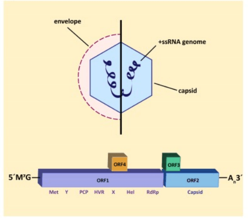
Schematic representation of hepatitis E virus particle (PMID: 31141919)
2.2 Hepatitis E Virus Genome & Classification
HEV belongs to the genus Orthohepevirus in the family Hepeviridae. Its genome is a positive‑sense single‑stranded RNA of approximately 7.2 kb.
Based on phylogenetic analysis, HEV is divided into 8 genotypes and various subtypes:
Genotype | Host range | Transmission route | Geographic distribution |
Genotype 1 (HEV-1) | Humans only | Faecal‑oral, waterborne | Resource‑limited countries |
Genotype 2 (HEV-2) | Humans only | Faecal‑oral, waterborne | Resource‑limited countries |
Genotype 3 (HEV-3) | Zoonotic | Consumption of undercooked pork, deer, rabbit, etc. | High‑income countries |
Genotype 4 (HEV-4) | Zoonotic | Consumption of undercooked animal meat | High‑income countries |
Genotype 7 (HEV-7) | Zoonotic | Animal contact/consumption | Limited distribution |
C1 (HEV-C1) | Zoonotic (rat HEV) | Contact with rats or their excreta | Global |
HEV classification includes two subfamilies: Orthohepevirinae and Parahepevirinae. HEV that infects humans belongs to the genus Paslahepevirus within the former.
2.3 What Are Hepatitis E Virus Structural Proteins?
The major structural and non‑structural proteins encoded by HEV are as follows:
Protein | Length (aa) | Main Function |
ORF1 polyprotein | ~1,693 | Non‑structural polyprotein; after proteolytic cleavage yields multiple functional domains including methyltransferase (Met), Y‑domain, papain‑like cysteine protease (PCP), hypervariable region (HVR), X‑domain, helicase (Hel), and RNA‑dependent RNA polymerase (RdRp) |
ORF2 (capsid protein) | ~660 | Major capsid structural protein; primary target of neutralizing antibodies; counteracts cell‑intrinsic antiviral responses to promote viral persistence |
ORF3 (VP13) | 113–114 | Multifunctional phosphoprotein; involved in viral release and mediates budding/secretion of quasi‑enveloped virions; exhibits prion‑like characteristics contributing to pathogenesis |
ORF4 | — | Present only in genotype 1; expressed under ER stress via IRES‑like mechanism; enhances viral replication |
2.3.1 ORF2 Capsid Protein
ORF2 is the major structural protein of HEV, forming the viral capsid. Recent studies have revealed that ORF2 plays multiple roles in the HEV life cycle: beyond its structural function, ORF2 counteracts cell‑intrinsic antiviral responses by interfering with antiviral signaling pathways and shielding viral replication from immune effectors, thereby promoting persistent HEV replication in immunocompetent cells.
2.3.2 ORF3 Multifunctional Protein
The phosphoprotein encoded by ORF3 (VP13) plays a critical role in viral release. Although HEV has traditionally been considered a non‑enveloped virus, recent findings show that it egresses from infected cells as quasi‑enveloped virions. ORF3 mediates sorting into vesicles and quasi‑enveloped virions through palmitoylation‑dependent association with Annexin II. A 2025 study published in PNAS further revealed that the prion‑like characteristic of ORF3 contributes to virion release and pathogenesis.
2.4 Mechanism of Cell Entry and Replication
2.4.1 Viral Attachment and Receptor Recognition
The cell entry mechanism of HEV is not fully elucidated. Known receptors and attachment factors include:
l Heparan sulfate proteoglycans (HSPGs) : involved in initial viral attachment
l Integrins: potentially involved in viral entry
l Other incompletely characterized receptor molecules
2.4.2 Viral Entry
HEV enters host cells via receptor‑mediated endocytosis. After entry, the viral genome is released into the cytoplasm. HEV can also infect intestinal cells—a 2025 study using human intestinal enteroids (HIEs) demonstrated that HEV predominantly infects proliferative transit‑amplifying cells, with rapid intestinal epithelial cell turnover facilitating efficient HEV replication and dissemination.
2.4.3 Viral RNA Replication
HEV replication occurs entirely within the host cell cytoplasm:
l ORF1 translation: upon entry, the positive‑sense genomic RNA is directly translated into the ORF1 non‑structural polyprotein
l Polyprotein processing: the ORF1 polyprotein is cleaved by proteases to release functional domains (Met, Y, PCP, HVR, Hel, RdRp, etc.)
l Replicase complex formation: ORF1 proteins assemble with genomic RNA and host factors to form the replicase complex
l RNA synthesis: negative‑strand RNA intermediates are synthesized using positive‑strand RNA as template, followed by synthesis of new positive‑strand genomic RNA and subgenomic RNA
ORF2 and ORF3 are translated from an approximately 2.2 kb subgenomic RNA generated during viral replication. ORF2 promotes persistent replication by counteracting cell‑intrinsic antiviral responses.
2.4.4 Viral Assembly and Release
Viral assembly and release involve the following steps:
l Capsid assembly: ORF2 capsid protein binds positive‑sense genomic RNA to form icosahedral capsids in the cytoplasm
l ORF3‑mediated release: ORF3 associates with Annexin II via palmitoylation‑dependent mechanisms, directing viral sorting into vesicles
l Quasi‑enveloped release: the virus is released from infected cells as quasi‑enveloped virions via budding
HEV, traditionally considered a non‑enveloped virus, actually exists in two forms: non‑enveloped virions (excreted in faeces, environmentally stable) and quasi‑enveloped virions (circulating in blood, involved in cell‑to‑cell transmission).
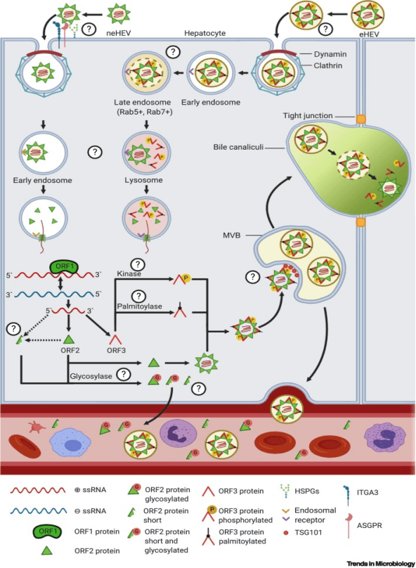
Schematic representation of the Hepatitis E Virus replication cycle and interaction with host factors.. (PMID: 32828646)
2.5 Symptoms and Treatment
2.5.1 Clinical Symptoms
The incubation period for HEV infection is typically 2–10 weeks. Approximately 70%–95% of HEV infections are asymptomatic or present with only mild symptoms. Common symptoms in symptomatic patients include:
l Nausea, vomiting
l Abdominal pain
l Jaundice
l Fever
l Malaise/fatigue
l Hepatomegaly
l Dark urine
High‑risk populations for severe disease:
l Pregnant women: significantly increased risk of fulminant hepatitis and death, with mortality rates up to 20%–30%
l Immunocompromised individuals (e.g., solid organ transplant recipients): can develop chronic HEV infection
l Patients with chronic liver disease: HEV superinfection significantly increases risks of liver failure and mortality
l Elderly: generally more severe disease course
Extrahepatic manifestations: HEV infection can also cause various extrahepatic manifestations, including neurological disorders (e.g., Guillain‑Barré syndrome) and renal injury.
2.5.2 Antiviral Treatment
Currently, no specific antiviral therapy is approved for HEV infection.
l Ribavirin: for fulminant hepatitis or chronic HEV infection, off‑label use of ribavirin is recommended. In immunocompromised patients, ribavirin combined with supportive care achieves significant clinical and laboratory improvement, resolving jaundice, restoring normal transaminase levels, and suppressing HEV RNA. Treatment of severe acute hepatitis E in immunocompetent patients typically uses ribavirin (400 mg every 12 hours). Ribavirin is contraindicated in pregnancy due to teratogenicity.
l Pegylated interferon alfa: alternative treatment option for patients with ribavirin‑treatment failure
l Sofosbuvir: shows inhibitory activity against HEV in cell culture models, but should not be used as monotherapy for HEV; no in‑vivo efficacy data currently available
l Bemnifosbuvir: a novel nucleotide analog showing additive antiviral effect when combined with ribavirin in chronic HEV infection
l Molnupiravir: shows anti‑HEV activity in animal models
2.6 Targets for Intervention
2.6.1 Viral Targets
Target | Protein | Biological Role |
ORF2 | Capsid protein | Virion structure, cell entry, immune antagonism |
RdRp (ORF1) | RNA‑dependent RNA polymerase | RNA replication |
ORF3 | Multifunctional phosphoprotein (VP13) | Viral release, quasi‑enveloped virion budding |
Helicase (ORF1) | Helicase | RNA unwinding |
Methyltransferase (ORF1) | Methyltransferase | RNA 5′ capping |
2.6.2 Host Receptors & Entry Factors
Host Target | Type | Role in Infection |
Heparan sulfate proteoglycans (HSPGs) | Attachment factors | Initial viral attachment |
Integrins | Receptors | Viral entry |
Proliferative intestinal stem cells/transit‑amplifying cells | Target cells | Intestinal HEV replication and dissemination |
2.6.3 Host Signaling Pathways
Pathway | Key Targets | Biological Role |
Interferon signaling | IFN‑α/β | Antiviral response, antagonized by ORF2 |
ER stress pathway | — | Triggers ORF4 expression (genotype 1) |
Cell‑intrinsic antiviral response | — | Suppressed by ORF2, promoting viral persistence |
2.7 Vaccine Types and Development Progress
Currently, only one HEV vaccine is licensed worldwide.
2.7.1 Hecolin® (HEV 239, Recombinant Protein Vaccine)
Hecolin® is a recombinant protein subunit vaccine developed by Xiamen Innovax Biotech Co., based on an E. coli‑expressed HEV ORF2 capsid protein fragment (aa 368–606). Key features:
l Recommended population: adults aged 16 years and older
l Standard regimen: 3 doses at 0, 1, and 6 months
l Licensed region: currently only in China
Key 2025 research advances:
l Two‑dose regimen effectiveness: a real‑world study during a HEV outbreak in the Bentiu refugee camp, South Sudan, demonstrated that a two‑dose Hecolin schedule can replace the three‑dose schedule during outbreak campaigns. Two‑dose vaccine effectiveness was 67.8%, increasing to 84.0% after confounder adjustment.
l Effectiveness in chronic hepatitis B patients: among HBsAg‑positive adults, HEV 239 vaccine effectiveness was 72.1% (95% CI 11.2–91.2), and 81.5% (95% CI 35.9–94.6) among Phase 3 trial participants.
l Immunogenicity and safety in chronic hepatitis B patients: in clinically stable CHB patients, anti‑HEV IgG seroconversion rates after three doses were >97%, with immunogenicity non‑inferior to healthy adults and no vaccine‑associated severe adverse events.
2.7.2 Other Vaccine Research
l Monoclonal antibody therapy: in 2025, the Pasteur Institute identified neutralizing human monoclonal antibodies effective against HEV, published in Science Advances. In another study, monoclonal antibody 5F6A1 significantly reduced viral load and viral shedding in infected pigs.
l Vaccine recommendations: the ESCMID Viral Hepatitis Study Group recommends HEV vaccination for high‑risk groups, including women of childbearing age.
l Vaccination during pregnancy: HEV vaccination during pregnancy may be feasible during outbreak settings.
2.8 Drugs
Currently, no specific small‑molecule antiviral is approved for HEV. Treatment relies primarily on supportive care and off‑label use.
2.8.1 Virus-Targeted Drugs
Target | Drug/candidate | Mechanism | Stage |
RdRp | Ribavirin | Nucleoside analog, RNA synthesis inhibition | Off‑label use |
Sofosbuvir | Nucleoside analog, RNA synthesis inhibition | Effective in vitro | |
Molnupiravir | Nucleoside analog, RNA synthesis inhibition | Animal models | |
Bemnifosbuvir | Nucleoside analog | Additive effect with ribavirin | |
Multiple targets | Pegylated interferon alfa | Immune modulation | Alternative after ribavirin failure |
ORF2 | Neutralizing mAbs (e.g., 5F6A1) | Viral neutralization | Preclinical |
2.8.2 Host-Targeted Drugs
Target | Drug | Mechanism | Stage |
Immunosuppression adjustment | — | Reduction of immunosuppressive drugs | First‑line strategy for chronic HEV in transplant recipients |
Supportive care | Electrolyte replacement, fluids | Supportive care | Standard of car |
2.8.3 Treatment Resistance
Ribavirin treatment failure is a significant clinical challenge. Ribavirin resistance‑associated mutations (e.g., V1479I) have been detected in clinical isolates. For patients with ribavirin treatment failure, alternative options including pegylated interferon alfa and sofosbuvir may be considered.
2.9 Epidemiology
HEV is a leading cause of acute viral hepatitis worldwide:
l Global infection burden: approximately 20 million HEV infections annually, with about 3.3 million symptomatic cases
l Mortality burden: approximately 19.5 million infections and 3,500 deaths globally in 2021
l Geographic distribution: HEV‑1 and HEV‑2 are endemic in resource‑limited countries, transmitted via faecal‑oral and waterborne routes; HEV‑3 and HEV‑4 are most common in high‑income countries, transmitted zoonotically
l Rat HEV: distantly related to HEV‑1 to HEV‑4, primarily distributed in rats, recently identified as a zoonotic pathogen capable of causing hepatitis in humans
2.10 Diagnosis
Diagnosis of HEV infection is recommended using a combination of serology and nucleic acid testing:
Test | Target | Clinical significance |
Anti‑HEV IgM | IgM antibodies | Marker of acute infection |
Anti‑HEV IgG | IgG antibodies | Past infection or vaccine response |
HEV RNA (RT‑PCR) | Viral RNA | Direct evidence of current infection |
HEV antigen detection | Viral antigen | Adjunctive diagnosis of current infection |
3. Related Products & Services
l Recombinant Antigens & Receptors
l Antibodies
l Detection Kits
l Recombinant Protein Expression Services
l Antibody Development Services
4. Resources
5. References
1. Kamar, N., Izopet, J., Pavio, N., et al. (2017). Hepatitis E virus infection. Nature Reviews Disease Primers, 3, 17086.
2. Nimgaonkar, I., Ding, Q., Schwartz, R. E., et al. (2018). Hepatitis E virus: advances and challenges. Nature Reviews Gastroenterology & Hepatology, 15(2), 96–110.
3. Brüggemann, Y., Klöhn, M., Wedemeyer, H., et al. (2024). Hepatitis E virus: from innate sensing to adaptive immune responses. Nature Reviews Gastroenterology & Hepatology, 21, 710–725.
4. Kamar, N., Bendall, R., Legrand-Abravanel, F., et al. (2012). Hepatitis E. The Lancet, 379(9835), 2477–2488.
5. Li, P., Liu, J., Li, Y., Su, J., Ma, Z., Bramer, W. M., Cao, W., de Man, R. A., Peppelenbosch, M. P., & Pan, Q. (2020). The global epidemiology of hepatitis E virus infection: A systematic review and meta-analysis. Liver International, 40(7), 1516–1528.
6. Montpellier, C., Wychowski, C., Sayed, I. M., et al. (2018). Hepatitis E virus lifecycle and identification of 3 forms of the ORF2 capsid protein. Gastroenterology, 154(1), 211–223.e8.
7. Yin, X., Ying, D., Lhomme, S., et al. (2018). Origin, antigenicity, and function of a secreted form of ORF2 in hepatitis E virus infection. Proceedings of the National Academy of Sciences of the United States of America, 115(18), 4773–4778.
8. Kenney, S. P., Meng, X. J. (2019). Hepatitis E virus genome structure and replication strategy. Cold Spring Harbor Perspectives in Medicine, 9(10), a031724.
9. Kanda, T., et al. (2024). Recent advances in hepatitis E virus research and the Japanese clinical practice guidelines for hepatitis E virus infection. Hepatology Research, 54.
10. Zhu, F. C., Zhang, J., Zhang, X. F., et al. (2010). Efficacy and safety of a recombinant hepatitis E vaccine in healthy adults: a large-scale, randomised, double-blind placebo-controlled, phase 3 trial. The Lancet, 376(9744), 895–902.
11. Shrestha, M. P., Scott, R. M., Joshi, D. M., et al. (2007). Safety and efficacy of a recombinant hepatitis E vaccine. The New England Journal of Medicine, 356(9), 895–903.
12. van der Valk, M., Zaaijer, H. L., Kater, A. P., et al. (2017). Sofosbuvir shows antiviral activity in a patient with chronic hepatitis E virus infection. Journal of Hepatology, 67(3), 666–669.
13. Pischke, S., Hartl, J., Pas, S. D., Lohse, A. W.,et al. (2017). Hepatitis E virus: infection beyond the liver? Journal of Hepatology, 66(5), 1082–1095.
14. Li, S., Li, W., Wang, X., et al. (2025). Integrin beta 1 facilitates non-enveloped hepatitis E virus cell entry through the recycling endosome. Nature Communications, 16.
15. Zhang, Y., et al. (2026). Broadly neutralizing antibodies isolated from HEV convalescents confer protective effects in human liver-chimeric mice. Nature Communications, 17.