
info@ucallmlabs.com
1 Overview
Influenza A virus (IAV) is the prototype species of the genus Influenza virus A within the family Orthomyxoviridae and is a major causative agent of seasonal and pandemic influenza in humans. It is an enveloped, negative‑sense, single‑stranded, segmented RNA virus with a pleiomorphic virion, approximately 50–120 nm in diameter.
Influenza A viruses are the only influenza viruses known to cause flu pandemics. Influenza A viruses infect multiple species, including humans, other mammals, and wild and domestic birds. Based on the antigenic differences of two major surface glycoproteins — hemagglutinin (HA) and neuraminidase (NA) — influenza A viruses are further divided into subtypes. Currently, 18 HA subtypes (H1–H18) and 11 NA subtypes (N1–N11) are known, with over 130 subtype combinations identified in nature. Current subtypes that routinely circulate in humans include A(H1N1) and A(H3N2). Influenza A viruses cause recurrent seasonal epidemics and occasional global pandemics with devastating levels of morbidity and mortality.
2.Background Information
2.1 What is Influenza A Virus?
Influenza A virus is an enveloped, negative-sense, single-stranded, segmented RNA virus belonging to the family Orthomyxoviridae and genus Influenzavirus A. The virion is pleiomorphic, approximately 50-120 nm in diameter. The viral envelope is derived from the host cell membrane and contains three major transmembrane proteins - hemagglutinin (HA), neuraminidase (NA), and matrix protein 2 (M2). The inner side of the envelope is lined by the matrix protein M1. The viral core consists of ribonucleoprotein complexes (vRNPs) containing eight segmented viral RNA molecules, each associated with nucleoprotein (NP) and the polymerase complex (PB1, PB2, PA).
The influenza A virus genome consists of 8 segmented negative‑sense RNA molecules with a total size of approximately 13 kb. The 8 genome segments encode 11 proteins:
| Segment | Protein | MW (kDa) | Major function |
| Segment 1 | PB2 | ~86 | Polymerase component, recognizes host cell RNA cap structure |
| Segment 2 | PB1 | ~86 | Polymerase catalytic subunit, nucleotide addition |
| Segment 2 (+1 frame) | PB1-F2 | ~11 | Pro‑apoptotic mitochondrial protein |
| Segment 3 | PA | ~81 | Polymerase component, possible transcriptase protease activity |
| Segment 4 | NP | ~62 | Nucleoprotein, binds viral RNA |
| Segment 6 | NA | ~52 | Neuraminidase, cleaves sialic acid, promotes virus release |
| Segment 7 | M1 | ~28 | Matrix protein, main component of viral capsid |
| Segment 7 (spliced) | M2 | ~11 | Ion channel protein |
| Segment 8 | NS1 | ~24 | Non‑structural protein, affects cellular RNA transport, splicing and translation |
| Segment 8 (+1 frame) | NEP/NS2 | ~13 | Nuclear export protein, mediates vRNP nuclear export |

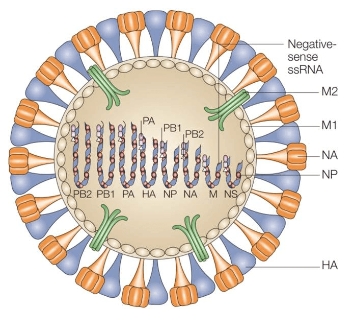
Schematic structure of an influenza A virus virion (PMID: 16064053)
2.2 Influenza A Virus Genome & Classification
Influenza A virus belongs to the genus Influenzavirus A in the family Orthomyxoviridae. Its genome is a negative‑sense single‑stranded RNA composed of 8 segments, listed from largest to smallest by convention. Each segment contains a coding region as well as short 5′ and 3′ flanking sequences.
Influenza A viruses are divided into subtypes based on the antigenic differences of two surface glycoproteins, HA and NA. Currently, 18 HA subtypes (H1–H18) and 11 NA subtypes (N1–N11) have been identified. The nomenclature for influenza virus isolates includes: influenza virus type (A), host species (omitted if human in origin), geographical site, serial number, year of isolation, and the H and N subtypes. For example, a human isolate is designated A/Brisbane/10/2007 (H3N2), while an avian isolate is A/duck/Hunan/795/2002 (H5N1).
Influenza A viruses have the ability to undergo "reassortment" - when two different subtypes infect a host simultaneously, they can swap gene segments. This is a key mechanism for the emergence of pandemic influenza strains.
2.3 What Are Influenza A Virus Structural Proteins?
Influenza A virus encodes 11 proteins, including multiple structural proteins and a few non‑structural proteins. The major structural proteins and their functions are as follows:
| Protein | Full name | Main Function |
| HA | Hemagglutinin | Trimeric surface glycoprotein, ~80% of envelope proteins; binds sialic acid receptors, mediates viral entry and membrane fusion |
| NA | Neuraminidase | Tetrameric surface glycoprotein; cleaves sialic acid, promotes release of new virions, prevents viral aggregation |
| M1 | Matrix protein 1 | Main component of viral capsid, lines inner envelope; involved in viral assembly |
| M2 | Matrix protein 2 | Ion channel protein; acidifies virion interior during uncoating, promotes RNP release |
| NP | Nucleoprotein | Binds tightly to viral RNA, forms ribonucleoprotein complexes |
| PB1/PB2/PA | Polymerase complex | Trimeric RNA‑dependent RNA polymerase, responsible for transcription and replication |
| NS1 | Non‑structural protein 1 | Non‑structural protein; affects cellular RNA transport, splicing and translation; antagonizes host interferon response |
| NEP/NS2 | Nuclear export protein | Mediates nuclear export of vRNPs |
2.4 Mechanism of Cell Entry and Replication
2.4.1 Viral Attachment and Receptor Recognition
Infection begins with the binding of viral HA protein to sialic acid (N-acetyl-neuraminic acid) receptors on host cell surfaces. HA contains two domains - HA1 and HA2. HA1 is responsible for receptor binding, while HA2 mediates membrane fusion. The presence and distribution of different types of sialic acid vary between humans, birds and other animals, which is a major determinant of host specificity.
Following HA‑sialic acid binding, the virus enters host cells via clathrin-dependent or ‑independent endocytosis or micropinocytosis.
2.4.2 Fusion and Uncoating
After endocytosis, endosome acidification triggers fusion of the viral and endosomal membranes. During this process, the M2 ion channel permits hydrogen ions to enter the virion, acidifying its interior and causing dissociation of M1 from vRNPs. This uncoating step allows vRNPs to be released into the cytoplasm and enter the nucleus. M2 channel inhibitors (such as amantadine and rimantadine) block this process.
2.4.3 Viral RNA Replication
A unique feature of influenza A virus is that it replicates within the host cell nucleus. After vRNPs enter the nucleus, the heterotrimeric polymerase complex (PA, PB1, PB2) and NP initiate viral RNA synthesis. The replication process includes:
Transcription of negative‑sense vRNA into mRNA, which is translated into viral proteins
Synthesis of positive‑sense cRNA intermediates using negative‑sense vRNA as template
Synthesis of progeny negative‑sense vRNA using positive‑sense cRNA as template
2.4.4 Viral Assembly and Release
Newly synthesized viral genomes (as vRNPs) are exported from the nucleus to the cytoplasm via NEP and M1 proteins. Assembly occurs at the plasma membrane in association with lipid rafts. HA, NA and M2 reach the plasma membrane via the Golgi apparatus, where M1 and vRNPs are incorporated into budding particles.
Efficient virus release requires NA to remove sialic acids from the cell surface or adjacent virions to prevent aggregation due to HA‑sialic acid binding. NA inhibitors (such as oseltamivir and zanamivir) inhibit NA activity and impair influenza virus release.
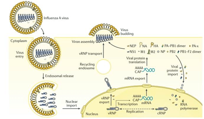
The life cycle of Influenza A virus. (PMID: 29955068)
2.5 Symptoms and Treatment
2.5.1 Clinical Symptoms
Influenza A virus infection can result in a broad clinical spectrum ranging from asymptomatic infection to severe pneumonia. Symptoms typically begin 1–4 days after infection. Common symptoms include:
Acute onset of fever
Cough (usually dry)
Sore throat
Body aches and fatigue
Headache
Muscle and joint pain
Runny nose
Most people recover within a week without requiring medical attention. However, severe cases may involve viral pneumonia, acute respiratory distress syndrome (ARDS), secondary bacterial pneumonia, and multi‑organ failure. The elderly, young children, pregnant women, and individuals with chronic underlying conditions are at higher risk for severe disease.
2.5.2 Antiviral Treatment
Antiviral treatment is most effective if initiated within 48 hours of symptom onset. For hospitalized patients, those with severe illness, or those at higher risk for complications, antiviral therapy may still be beneficial if started >48 hours after illness onset.
Four antiviral agents approved by the U.S. FDA for the treatment and prophylaxis of influenza are available:
Oseltamivir (Tamiflu®) : oral administration, recommended for all ages; preferred agent for hospitalized patients, severe or complicated influenza
Zanamivir (Relenza®) : inhaled administration, approved for treatment in patients ≥7 years and prophylaxis in people ≥5 years
Peramivir (Rapivab®) : intravenous administration, approved for patients ≥2 years; useful for those unable to tolerate oral therapy
Baloxavir (Xofluza®) : oral single‑dose, indicated for acute uncomplicated influenza in otherwise healthy patients ≥5 years and high‑risk patients ≥12 years
Two other FDA‑approved drugs — amantadine and rimantadine — are not recommended for treatment or prophylaxis due to widespread viral resistance.
WHO conditionally recommends treatment with oseltamivir within 48 hours of symptom onset for severe illness, and baloxavir for patients at high risk of progression from non‑severe to severe illness.
2.6 Targets for Intervention
2.6.1 Viral Targets
| Target | Protein | Biological Role |
| HA | Hemagglutinin | Receptor binding & membrane fusion |
| NA | Neuraminidase | Cleaves sialic acid, promotes virus release |
| M2 | Ion channel | Viral uncoating |
| Polymerase complex | PB1/PB2/PA | RNA transcription & replication |
| NP | Nucleoprotein | RNA binding & vRNP formation |
2.6.2 Host Receptors & Entry Factors
| Host Target | Type | Role in Infection |
| Sialic acid receptor | Receptor | HA‑mediated viral attachment |
| Clathrin‑mediated endocytosis | Endocytic pathway | Viral entry |
| Host proteases | Protease | HA proteolytic cleavage activation |
2.6.3 Host Signaling Pathways
| Pathway | Key Targets | Biological Role |
| RIG‑I signaling | RIG‑I | Viral RNA sensing, inhibited by NS1 |
| Interferon signaling | IFN‑α/β | Antiviral response |
| NF‑κB pathway | NF‑κB | Inflammatory response |
| JAK–STAT pathway | JAK1, STAT1/2 | Interferon signaling |
2.7 Vaccine Types and Development Progress
Influenza vaccines are the most effective measure for preventing influenza A virus infection. Due to continuous antigenic drift of influenza viruses, vaccine components must be updated annually based on global surveillance data.
2.7.1 Conventional Inactivated Vaccines (IIV)
The first inactivated influenza vaccines were developed by growing large quantities of influenza viruses in embryonated hens' eggs, collecting virus‑containing allantoic fluid, and then purifying and inactivating the whole virus. Modern inactivated vaccines include whole‑virus inactivated, split‑virus, and subunit vaccines.
2.7.2 Live Attenuated Vaccines (LAIV)
Intranasally administered live attenuated influenza vaccines induce mucosal immunity in the upper respiratory tract, mimicking natural infection
2.7.3 Recombinant HA Vaccines
HA protein vaccines produced using recombinant DNA technology do not rely on egg‑based production and can shorten the vaccine manufacturing timeline.
2.7.4 mRNA Vaccines
The mRNA platform offers potential benefits for influenza vaccine production. Moderna's mRNA‑1010 quadrivalent influenza vaccine (containing HA of influenza A H1N1, H3N2, and both B lineages) has entered Phase 3 clinical trials. Preliminary results demonstrate comparable immunogenicity to COVID‑19 vaccines and more robust immunogenicity against influenza A.
2.7.5 Universal Influenza Vaccines
Universal influenza vaccines represent a frontier in influenza vaccine development, aiming to provide broad protection against all influenza virus strains (including seasonal variants and potential pandemic strains). Major strategies include:
Targeting conserved HA stalk domain: chimeric HA (cHA)‑based vaccines induce cross‑reactive antibodies targeting the HA stalk
Targeting conserved antigens (NP, M2e) : mucosal immunization with conserved NP antigens induces broadly protective immune responses against both influenza A and B viruses
Nanoparticle vaccines: e.g., structure‑guided HA stem nanoparticle vaccine (SteMos1) has entered Phase I clinical trials
Recombinant NA protein vaccines: e.g., rNA‑N2‑MPP and other candidates have shown broad protection potential in preclinical studies
Multiple universal influenza vaccine candidates have entered clinical trials, including Osivax's OVX836 (7 clinical trials with over 1,400 participants), Centivax's Centi‑Flu 01 (pan‑influenza vaccine, Phase I initiated), and others.
2.8 Drugs
Antiviral drugs for influenza A virus can be classified by their target as follows:
2.8.1 Virus-Targeted Drugs
| Target | Drug/candidate | Mechanism |
| NA | Oseltamivir | Neuraminidase inhibition, prevents virus release |
| Zanamivir | Neuraminidase inhibition | |
| Peramivir | Neuraminidase inhibition | |
| Polymerase (PA) | Baloxavir | Cap‑dependent endonuclease inhibition, blocks viral replication |
| M2 ion channel | Amantadine | M2 channel blockade |
| Rimantadine | M2 channel blockade |
2.8.2 Host-Targeted Drugs
| Target | Drug/candidate | Mechanism |
| Host inflammation | Corticosteroids | Immunosuppression |
| JAK‑STAT pathway | Baricitinib | JAK inhibition |
2.8.3 Drug Resistance
Antiviral drug resistance in influenza viruses requires ongoing surveillance. Between July and October 2025, 20% to 100% per week of influenza A(H1N1)pdm09 strains characterized in Catalonia, Spain, carried the NA:S247N substitution, which conferred reduced susceptibility to oseltamivir. Additionally, spontaneous emergence of drug‑resistant highly pathogenic avian influenza A(H5N1) viruses poses a persistent threat to human health. WHO global surveillance data indicate that resistance mutations to baloxavir in seasonal influenza viruses have remained at extremely low levels from 2018 to 2025.
3 Related Products & Services
Recombinant Antigens & Receptors
Antibodies
Detection Kits
Recombinant Protein Expression Services
Antibody Development Services
4 Resources
5 References
Krammer, F., Smith, G. J. D., Fouchier, R. A. M., et al. (2018). Influenza. Nature Reviews Disease Primers, 4, 3.
Neumann, G., Cowling, B. J., Chen, H., et al. (2026). Influenza. Nature Reviews Disease Primers, 12, 51.
Bouvier, N. M., & Palese, P. (2008). The biology of influenza viruses. Vaccine, 26(Suppl 4), D49–D53.
Dadonaite, B., Gilbertson, B., Knight, M. L., et al. (2019). The structure of the influenza A virus genome. Nature Microbiology, 4, 1781–1789.
te Velthuis, A. J. W., & Fodor, E. (2016). Influenza virus RNA polymerase: Insights into the mechanisms of viral RNA synthesis. Nature Reviews Microbiology, 14, 479–493.
Eisfeld, A. J., Neumann, G., & Kawaoka, Y. (2015). At the centre: Influenza A virus ribonucleoproteins. Nature Reviews Microbiology, 13, 28–41.
Shi, Y., Wu, Y., Zhang, W., Qi, J., & Gao, G. F. (2014). Enabling the 'host jump': Structural determinants of receptor-binding specificity in influenza A viruses. Nature Reviews Microbiology, 12, 822–831.
Long, J. S., Mistry, B., Haslam, S. M., & Barclay, W. S. (2019). Host and viral determinants of influenza A virus species specificity. Nature Reviews Microbiology, 17, 67–81.
Pflug, A., Guilligay, D., Reich, S., et al. (2014). Structure of influenza A polymerase bound to the viral RNA promoter. Nature, 516, 355–360.
Rossman, J. S., & Lamb, R. A. (2011). Influenza virus assembly and budding. Virology, 411, 229–236.
Nayak, D. P., Balogun, R. A., Yamada, H., Zhou, Z. H., & Barman, S. (2009). Influenza virus morphogenesis and budding. Virus Research, 143, 147–161.
Krammer, F., & Palese, P. (2015). Advances in the development of influenza virus vaccines. Nature Reviews Drug Discovery, 14, 167–182.
Nachbagauer, R., & Palese, P. (2020). Is a universal influenza virus vaccine possible? Annual Review of Medicine, 71, 315–327.
Soens, M., Ananworanich, J., Hicks, B., et al. (2025). A phase 3 randomized safety and immunogenicity trial of mRNA-1010 seasonal influenza vaccine in adults. Vaccine, 50, 126847.
Uyeki, T. M., Hui, D. S., Zambon, M., Wentworth, D. E., & Monto, A. S. (2022). Influenza. The Lancet, 400(10353), 693–706.
Kumari, R., Sharma, S. D., Kumar, A., et al. (2023). Antiviral Approaches against Influenza Virus. Clinical Microbiology Reviews, 36, e00040-22.
Beigel, J. H., & Hayden, F. G. (2021). Influenza Therapeutics in Clinical Practice—Challenges and Recent Advances. Cold Spring Harbor Perspectives in Medicine, 11, a038463.
Uehara, T., Hayden, F. G., Kawaguchi, K., et al. (2020). Treatment-emergent influenza variant viruses with reduced baloxavir susceptibility: Impact on clinical and virologic outcomes in uncomplicated influenza. Journal of Infectious Diseases, 221, 346–355.
Bright, R. A., Medina, M.-J., Xu, X., et al. (2005). Incidence of adamantane resistance among influenza A (H3N2) viruses isolated worldwide from 1994 to 2005: A cause for concern. The Lancet, 366, 1175–1181.
Saubi, N., Andrés, C., Prats-Méndez, I., et al. (2025). Expansion of influenza A(H1N1)pdm09 NA:S247N viruses with reduced susceptibility to oseltamivir, Catalonia, Spain, and in Europe, July to October 2025. Eurosurveillance, 30(48), 2500873.
Hussain, S., Meijer, A., Govorkova, E. A., et al. (2025). Global update on the susceptibilities of influenza viruses to neuraminidase inhibitors and the cap-dependent endonuclease inhibitor baloxavir, 2020–2023. Antiviral Research, 241, 106217.
World Health Organization. (2024). Clinical practice guidelines for influenza. Geneva: World Health Organization.
World Health Organization. (2026). Laboratory methodologies for testing the antiviral susceptibility of influenza viruses: Polymerase acidic (PA) protein inhibitor, baloxavir.
Kikkert, M. (2020). Innate immune evasion by human respiratory RNA viruses. Journal of Innate Immunity, 12, 4–20.
Ayllón, J., & García-Sastre, A. (2015). The influenza A virus NS1 protein: A multifaceted virulence factor. Current Topics in Microbiology and Immunology, 386, 1–41.