
info@ucallmlabs.com
1.Overview
Rabies virus (RABV) is the prototype species of the genus Lyssavirus within the family Rhabdoviridae and is the etiological agent of rabies. It is an enveloped, negative‑sense, single‑stranded RNA virus with a bullet‑shaped virion, approximately 130-250 nm in length and 60-110 nm in diameter. Rabies is an ancient zoonotic disease that, once clinical symptoms appear, is nearly 100% fatal - the highest case‑fatality rate among all viral infections. Transmission to humans occurs primarily through bites or scratches from infected animals via contaminated saliva. Globally, approximately 59,000 people die from rabies each year, with about 95% of deaths occurring in remote rural areas of Asia and Africa. Dogs are the most important reservoir host in Asia and are responsible for the majority of human rabies cases; worldwide, bats (Chiroptera) serve as the main reservoir for most lyssaviruses.
2.Background Information
2.1 What is Lyssavirus Rabies?
Rabies lyssavirus is an enveloped, negative‑sense, single‑stranded RNA virus belonging to the family Rhabdoviridae and genus Lyssavirus. The virion is bullet‑shaped or bacilliform, composed of two structural units: an internal helical nucleocapsid (diameter ~50 nm) and a lipid envelope derived from the host cell plasma membrane. The envelope surface is covered with spikes formed by trimers of the glycoprotein (G), each about 8 nm in length.
The viral genome is a negative‑sense, single‑stranded RNA of approximately 11.9-12.3 kb. The genome encodes five structural proteins in the order 3′‑N‑P‑M‑G‑L‑5′:
Nucleoprotein (N) : 58–62 kDa, tightly binds viral RNA and forms the nucleocapsid core.
Phosphoprotein (P) : 35–40 kDa, polymerase cofactor involved in transcription and replication, and suppresses host interferon responses.
Matrix protein (M) : 22–25 kDa, located on the inner side of the envelope, links the nucleocapsid to the envelope and participates in assembly and budding.
Glycoprotein (G) : 65–80 kDa, forms surface spikes, mediates receptor binding and membrane fusion, and is the primary target of neutralizing antibodies.
Large protein (L) : Mr ~190 kDa, the RNA‑dependent RNA polymerase (RdRp) responsible for transcription and replication of the viral genome.

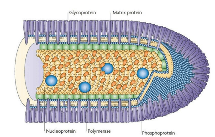
Schematic structure of rabies virion (PMID: 21601039)
2.2 Lyssavirus Rabies Genome & Classification
Rabies virus belongs to the genus Lyssavirus, whose members possess a negative‑sense single‑stranded RNA genome of about 12 kb. The genome organization is simple, containing only five open reading frames (ORFs) in the order 3′‑N‑P‑M‑G‑L‑5′. Both 3′ and 5′ ends contain untranslated regions (UTRs), with a longer 3′ UTR following the G mRNA.
The genus Lyssavirus exhibits genetic and antigenic diversity, with greater variability in the glycoprotein (G) than in the nucleoprotein (N). Different viral variants are often associated with specific host species. Phylogenetic analysis reveals that the genus comprises multiple virus species distributed worldwide (except Antarctica and some isolated islands).
2.3 What Are Lyssavirus Rabies Structural Proteins?
Rabies virus encodes five major structural proteins:
| Protein | Full name | Main Function |
| N | Nucleoprotein | Binds tightly to viral RNA, forms the nucleocapsid core, protects genomic RNA |
| P | Phosphoprotein | Polymerase cofactor; involved in transcription/replication; suppresses host interferon responses |
| M | Matrix protein | Inner envelope protein; links nucleocapsid to envelope; mediates assembly and budding |
| G | Glycoprotein | Surface spikes; mediates receptor binding and membrane fusion; primary target of neutralizing antibodies |
| L | Large protein | RNA‑dependent RNA polymerase (RdRp); catalyzes transcription and replication |
In addition, both N and P are phosphoproteins – N inhibits RIG‑I‑mediated type I interferon activation, while P expresses multiple isoforms (P1–P5) that suppress interferon induction and interferon‑dependent signaling. M protein is not phosphorylated; it interacts with RNP and G during assembly to form bullet‑shaped virions.
2.4 Mechanism of Cell Entry and Replication
2.4.1 Viral Attachment and Receptor Recognition
Infection begins with the binding of G protein on the virion surface to host cell receptors. Several receptor molecules have been identified to participate in rabies virus attachment and entry, including:
Nicotinic acetylcholine receptor (nAChR) : located at neuromuscular junctions, may concentrate virus at nerve endings.
Integrin β1 (ITGB1) : can enhance viral entry in human cells.
Other incompletely characterized receptor molecules – entry involves multiple receptors acting cooperatively.
Virus enters host cells via receptor‑mediated endocytosis, followed by clathrin‑mediated endocytosis and low‑pH‑dependent membrane fusion to complete uncoating.
2.4.2 Viral Entry and Neural Spread
A key feature of rabies virus infection is its neurotropism. After entering the host at the bite site, the virus can directly enter peripheral nerve axons and be transported retrogradely to the central nervous system (CNS). It may also replicate locally in muscle tissue at the bite site, persist near the site during the incubation period, and then invade the nervous system and spread to the brain. Once reaching the CNS, it causes acute encephalomyelitis and then spreads centrifugally to multiple organs.
2.4.3 Viral RNA Replication
After entry and uncoating, the L protein (RdRp) carried by the virion initiates transcription of the genome. Replication involves:
Transcription of the negative‑sense RNA genome into mRNAs, which are translated into five structural proteins.
During replication, positive‑sense RNA intermediates are synthesized using the negative‑sense RNA as template.
Progeny negative‑sense RNA genomes are synthesized using positive‑sense RNA as template.
Newly synthesized N protein immediately binds to progeny negative‑sense RNA to form ribonucleoprotein complexes (RNPs).
2.4.4 Viral Assembly and Release
Assembly begins with the interaction of RNP with M protein. M protein connects RNP to regions of the cell membrane enriched with G protein. The virus acquires its envelope by budding from the host cell membrane, forming bullet‑shaped virions that are released.
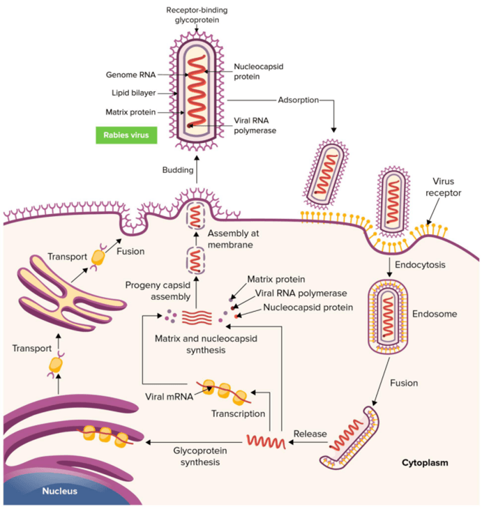
The life cycle of rabies virus. (PMID: 19946287)
2.5 Symptoms and Treatment
2.5.1 Clinical Symptoms
The clinical presentation of rabies can be divided into five stages: incubation, prodrome, acute neurological phase, coma, and death (with rare recovery).
Incubation period: highly variable, ranging from less than 10 days to over 2 years, typically 1–3 months. Duration depends on bite site, viral load, and host immune status.
Prodrome: usually lasts 2–10 days, with non‑specific symptoms including malaise, fever, fatigue, sore throat, cough, dyspnoea, anorexia, dysphagia, nausea, vomiting, and abdominal pain.
Acute neurological phase: mainly two clinical forms:
Furious (encephalitic) rabies: most common, characterised by agitation, aggressive behaviour, hydrophobia, aerophobia, and autonomic dysfunction.
Paralytic (dumb) rabies: progressive muscle weakness and paralysis, usually starting near the bite site.
Once clinical symptoms appear, rabies is almost invariably fatal.
2.5.2 Post-Exposure Prophylaxis (PEP)
PEP is currently the only effective intervention and includes:
Wound care - immediate and thorough washing and disinfection of the wound.
Passive immunization - infiltration of human rabies immunoglobulin (HRIG) around the wound.
Active immunization - administration of rabies vaccine.
Traditional PEP regimens include the 5‑dose Essen regimen or the 4‑dose Zagreb regimen. Timely and correct PEP effectively prevents disease progression. However, multiple doses and high costs impose a heavy burden in resource‑limited settings. No effective antiviral therapy exists once clinical symptoms appear.
2.6 Targets for Intervention
2.6.1 Viral Targets
| Target | Protein | Biological Role |
| G protein | Glycoprotein (G) | Receptor binding & membrane fusion |
| L protein | RNA polymerase (L) | RNA synthesis |
| N protein | Nucleoprotein (N) | RNA binding & genome packaging |
| P protein | Phosphoprotein (P) | Transcription/replication, interferon antagonism |
| M protein | Matrix protein (M) | Assembly & budding |
2.6.2 Host Receptors & Entry Factors
| Host Target | Type | Role in Infection |
| nAChR | Receptor | Virus attachment, enrichment at nerve endings |
| ITGB1 | Receptor | Enhances viral entry |
| Clathrin‑mediated endocytosis | Endocytic pathway | Viral entry |
2.6.3 Host Signaling Pathways
| Pathway | Key Targets | Biological Role |
| RIG‑I signaling | RIG‑I | Viral RNA sensing, inhibited by N protein |
| Interferon signaling | IFN‑α/β | Antiviral response, suppressed by P protein |
| Nrf2/SQSTM1/PINK1/Parkin | Nrf2, SQSTM1 | Oxidative stress and autophagy pathways |
2.7 Vaccine Types and Development Progress
Rabies vaccines have evolved from nerve‑tissue vaccines to modern cell‑culture vaccines. Several platforms are available, including inactivated, live‑attenuated, recombinant, viral‑vectored, and nucleic‑acid vaccines.
2.7.1 Conventional Inactivated Vaccines (Cell-Culture)
Human diploid cell vaccine (HDCV) e.g., Lyssavac‑HDC - good immunogenicity and safety.
Purified Vero cell vaccine (PVRV) - second‑generation human vaccine, still requires 5- or 4-dose regimens.
Purified duck embryo vaccine (PDEV) e.g., Lyssavac‑N - can be used in WHO‑approved intradermal PEP schedules.
2.7.2 mRNA Vaccines
mRNA rabies vaccines represent a promising next‑generation approach. Studies show:
Full‑length RABV-G mRNA vaccines induce strong immune responses and provide complete protection.
RABV‑Full and RABV-R333Q mRNA vaccines perform best, while those encoding only ectodomain or transmembrane domain are less effective.
Dual‑antigen mRNA vaccine (RABV-G-LT) provides 100% protection in mice, superior to inactivated vaccine.
2.7.3 DNA vaccines
DNA vaccines are another novel platform. Research directions include optimizing antigen constructs and heterologous prime‑boost strategies.
2.7.4 Viral-Vectored Vaccines
Rabies virus itself is used as a vaccine vector. Three types of RV‑vectored vaccines exist: live‑attenuated, inactivated, and replication‑deficient vectors.
2.7.5 Novel Vaccine Directions
Single‑dose vaccines - reduce the number of doses and economic burden.
Oral vaccines - gut-targeted oral vaccines for humans are a promising future direction.
Combination vaccines - e.g., Lassa‑rabies bivalent vaccine has shown safety and ability to elicit antibody responses against both viruses in early clinical trials.
Despite effective vaccine prevention, rabies remains a major global disease burden, mainly due to insufficient vaccination coverage, especially in resource‑limited regions.
2.8 Drugs
Currently, no specific antiviral drug is licensed for rabies. Once clinical symptoms appear, existing antivirals are ineffective. However, recent progress has been made in anti‑rabies drug discovery, classified by target as follows:
2.8.1 Virus-Targeted Drugs
| Target | Drug/candidate | Mechanism |
| Viral RNA G‑quadruplex (G4) | Λ‑M10 (metallo‑helical compound) | Stabilizes G4, inhibits G translation and replication |
| P protein‑LC8 interaction | Pep2 (peptide) | Inhibits protein‑protein interaction |
| Viral replication | BCX4430 | Inhibits mTOR‑dependent autophagy |
| Favipiravir (T705) | RNA synthesis inhibition | |
| Ribonucleoside analog (DON) | RNA synthesis inhibition |
2.8.2 Host-Targeted Drugs
| Target | Drug/candidate | Mechanism |
| Nrf2/SQSTM1/PINK1/Parkin | MG132 (proteasome inhibitor) | Inhibits RABV replication |
| Multiple kinases | Sunitinib | Protease inhibition |
| Artemisinin derivatives | Artesunate (ART), Dihydroartemisinin (DHA) | Antiviral activity |
2.8.3 Drugs with Unclear or Non-Specific Targets
| Drug | Mechanism | Stage |
| Ribavirin | Broad‑spectrum antiviral, unclear mechanism | In vitro |
| FDA‑approved drugs from high‑throughput phenotypic screening | Reduce RABV replication at low concentrations | Preliminary screening |
| AI‑designed candidate compounds | AI‑assisted anti‑RABV drug design | Computational design |
3.Related Products & Services
Recombinant Antigens & Receptors
Antibodies
Detection Kits
Recombinant Protein Expression Services
Antibody Development Services
4.Resources
5.References
International Committee on Taxonomy of Viruses. (2024). Genus: Lyssavirus. In ICTV Online (10th) Report. International Committee on Taxonomy of Viruses.
Hooper, D. C. (2016). Rabies virus. In D. D. Richman, R. J. Whitley, & F. G. Hayden (Eds.), Manual of molecular and clinical laboratory immunology (8th ed., pp. 665–673). ASM Press.
Finke, S., Conzelmann, K.-K. (2005). Replication strategies of rabies virus. Virus Research, 111(2), 120–131.
Rupprecht, C. E. (2011). Rhabdoviruses: Rabies virus. In S. Baron (Ed.), Medical microbiology (4th ed.). University of Texas Medical Branch at Galveston.
Schnell, M. J., McGettigan, J. P., Peltzer, M. K., et al. (2010). The cell biology of rabies virus: Using stealth to reach the brain. Nature Reviews Microbiology, 8, 51–61.
Yuan, S., Zhang, Z.-W., Jiang, S.-C., Li, Z.-L., & Hu, J. (2026). New treatments to rabies virus infections. Antiviral Research, 248, 106380.
Zhang, X., Qin, G., Lv, S., et al. (2026). Exploring RNA G-quadruplex in the rabies virus genome and its potential against RABV infection. Chemical Science, 17(2), 1232–1241.
Chi, Y. L., Yang, N., Xie, Y., et al. (2026). MG132-mediated inhibition of rabies virus replication via the Nrf2/SQSTM1/PINK1/Parkin autophagy pathway. Virology Journal, 23(1), 176.
Natesan, K., Isloor, S., Vinayagamurthy, B., et al. (2023). Developments in rabies vaccines: The path traversed from Pasteur to the modern era of immunization. Vaccines, 11(4), 756.
Rahmati, S., Zandi, F., Ahmadi, K., et al. (2025). Computational structure-based design of antiviral peptides as potential protein–protein interaction inhibitors of rabies virus phosphoprotein and human LC8. Heliyon, 11(1), e41520.
Xu, H., Hao, X., Wang, S., et al. (2015). Real-time imaging of rabies virus entry into living Vero cells. Scientific Reports, 5, 11753.