
info@ucallmlabs.com
Chondrocytes are the key functional cells responsible for maintaining the structure and function of articular cartilage. They synthesize cartilage-specific extracellular matrix (ECM) components, including type II collagen and aggrecan, which are essential for cartilage integrity and mechanical properties. Mesenchymal Stem Cells (MSCs) possess self-renewal capacity and multilineage differentiation potential, and under specific induction conditions, they can recapitulate embryonic chondrogenesis processes. Through sequential stages including mesenchymal condensation, chondrogenic lineage commitment, chondrocyte maturation, and extracellular matrix formation, MSCs can differentiate into chondrocytes with typical cartilage phenotypes.
In vitro MSC chondrogenic differentiation systems commonly utilize high-density three-dimensional pellet culture combined with chondrogenic induction medium. The chondrogenic process is regulated by coordinated stimulation of growth factors from the TGF-β family (particularly TGF-β3) and BMP family members such as BMP-2, BMP-6, and BMP-7. Activation of the TGF-β/Smad signaling pathway promotes the expression of the key transcription factor SOX9, which subsequently drives the expression of cartilage-specific genes including COL2A1 and ACAN, facilitating type II collagen and proteoglycan deposition and the formation of mature cartilage-like extracellular matrix.
During differentiation, MSCs first transition into chondrogenic progenitor cells, characterized by the expression of key cartilage lineage-determining transcription factors such as SOX9, SOX5, and SOX6. Subsequently, cells gradually acquire mature chondrocyte characteristics, exhibiting increased expression of cartilage matrix markers including COL2A1, ACAN, and COMP, accompanied by glycosaminoglycan (GAG) accumulation and cartilage-like tissue formation. By optimizing the culture environment, including hypoxic conditions, three-dimensional scaffolds, and biomaterial-based systems, the efficiency of MSC chondrogenic differentiation can be further enhanced while maintaining a stable hyaline cartilage phenotype.
MSC-derived chondrocyte systems provide important cellular models for cartilage repair studies, osteoarthritis mechanism research, tissue engineering, biomaterial evaluation, and drug screening. Due to their broad tissue sources, strong expansion capacity, and potential for engineering-based regulation, MSC-derived chondrocytes have become an important technological platform in regenerative medicine for investigating cartilage formation, functional reconstruction, and therapeutic development.
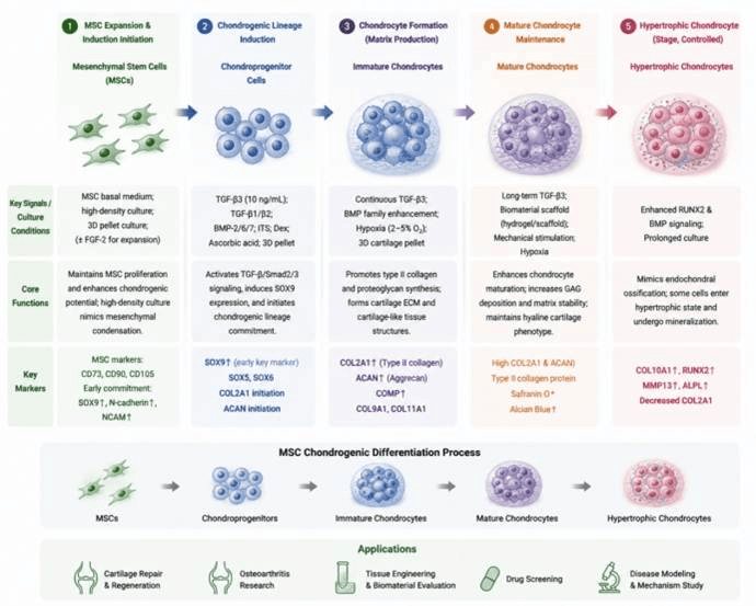
Mesenchymal stem cells (MSCs) undergo stepwise chondrogenic induction in vitro and progressively differentiate into chondroprogenitor cells, immature chondrocytes, and mature chondrocytes, forming cartilage-like extracellular matrix enriched in type II collagen and proteoglycans. This system recapitulates in vivo cartilage development and provides a stable cellular model for cartilage regeneration, osteoarthritis research, tissue engineering, and drug screening.
| Differentiation Stage | Added Factors / Culture Conditions | Core Biological Functions | Corresponding Cell Type / Developmental Stage | Stage-Specific Identification Markers |
| MSC Expansion and Chondrogenic Induction Initiation Stage | MSC basal culture system; high-density culture; 3D pellet culture; some systems include FGF-2 to enhance expansion | Maintains MSC proliferation capacity and enhances chondrogenic differentiation potential; high-density culture mimics mesenchymal condensation during embryonic cartilage development | Undifferentiated Mesenchymal Stem Cells (MSCs) → Early Chondroprogenitors | MSC markers: CD73, CD90, CD105; Early chondrogenic commitment: SOX9↑, N-cadherin↑, NCAM↑ |
| Chondrogenic Lineage Induction Stage (Early Chondrogenic Stage) | TGF-β3 (commonly 10 ng/mL); TGF-β1/TGF-β2; BMP-2, BMP-6, BMP-7; ITS supplement; Dexamethasone (Dex); Ascorbic acid; 3D pellet culture | Activates TGF-β/Smad2/3 signaling, induces SOX9 expression, initiates cartilage-specific gene transcription, and promotes MSC transition toward chondrogenic progenitors | Chondroprogenitor Cells | SOX9↑ (key early marker); SOX5, SOX6; initiation of COL2A1 expression; onset of ACAN expression |
| Chondrocyte Formation Stage (Mature Cartilage Matrix Production Stage) | Continuous TGF-β3 stimulation; BMP family enhancement; hypoxic culture (2–5% O₂) to maintain cartilage phenotype; 3D cartilage pellet culture | Promotes type II collagen and proteoglycan synthesis, formation of cartilage extracellular matrix (ECM), and development of cartilage-like tissue structures | Immature Chondrocytes | COL2A1↑ (Type II collagen); ACAN↑ (Aggrecan); COMP↑; COL9A1, COL11A1 |
| Mature Chondrocyte Maintenance Stage | Long-term chondrogenic induction culture; TGF-β3 maintenance; biomaterial scaffolds (hydrogel/scaffold); mechanical stimulation | Promotes chondrocyte maturation, enhances glycosaminoglycan (GAG) deposition and cartilage matrix stability, and maintains the hyaline cartilage phenotype | Mature Chondrocytes | Sustained high expression of COL2A1 and ACAN; Type II collagen protein expression; Safranin O positive; Alcian Blue positive |
| Hypertrophic Chondrocyte Stage (Controlled Terminal Stage) | Enhanced RUNX2 and BMP signaling; prolonged culture may naturally induce hypertrophic differentiation | Mimics the process of endochondral ossification; some MSC-derived chondrocytes may enter a hypertrophic state and undergo mineralization | Hypertrophic Chondrocytes | COL10A1↑, RUNX2↑, MMP13↑, ALPL↑; decreased COL2A1 expression |