
info@ucallmlabs.com
Mesenchymal Stem Cells (MSCs) are adult stem cells with self-renewal capacity and multilineage differentiation potential. Under specific induction conditions, MSCs can undergo directed differentiation into osteoblasts, making them an important cell source for bone tissue engineering, bone regenerative medicine, and disease modeling studies. Classical studies have demonstrated that MSCs derived from various tissues, including bone marrow, adipose tissue, and umbilical cord, possess osteogenic differentiation potential. Following in vitro osteogenic induction, MSCs gradually progress through multiple developmental stages, including osteogenic lineage commitment, pre-osteoblast formation, mature osteoblast differentiation, and extracellular matrix mineralization, ultimately generating functional osteoblasts with bone matrix synthesis and mineralization capabilities.
The differentiation of MSCs into osteoblasts is precisely regulated by multiple signaling pathways, among which RUNX2 (Runt-related transcription factor 2) and SP7/Osterix serve as key transcriptional regulators determining osteogenic lineage commitment. Classical osteogenic induction systems typically utilize a combination of dexamethasone (Dex), ascorbic acid (Asc), and β-glycerophosphate (β-GP). Dex promotes the expression of osteogenic genes such as RUNX2, Asc facilitates type I collagen (COL1A1) synthesis and extracellular matrix formation, while β-GP provides a phosphate source for hydroxyapatite deposition and promotes late-stage bone matrix mineralization.
During the early stage of differentiation, MSCs gradually acquire osteoprogenitor characteristics, accompanied by increased expression of RUNX2, SP7, and alkaline phosphatase (ALP). Subsequently, cells enter the mature osteoblast stage, continuously producing bone matrix proteins including type I collagen, osteopontin (OPN), and bone sialoprotein (BSP). At the terminal differentiation stage, mature osteoblasts expressing osteocalcin (OCN) are generated and exhibit strong mineralization capacity through calcium deposition and hydroxyapatite formation. Alizarin Red S and Von Kossa staining are commonly used to evaluate the mineralization level and functional maturation of MSC-derived osteoblasts.
With the development of optimized MSC osteogenic induction technologies, MSC-derived osteoblasts have been widely applied in studies of bone formation mechanisms, evaluation of bone defect repair, screening of bone tissue engineering materials, drug toxicity assessment, and modeling of bone metabolism-related diseases. By integrating cellular morphology analysis, osteogenic gene expression profiling, protein marker detection, and mineralization functional assays, the directed differentiation process from MSCs to functional osteoblasts can be systematically validated, providing reliable cellular models for bone regeneration research and translational applications.
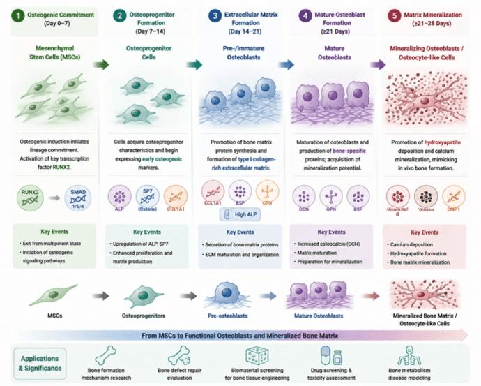
Mesenchymal stem cells (MSCs) undergo stepwise osteogenic induction and progressively differentiate into osteoprogenitor cells, mature osteoblasts, and ultimately mineralized bone matrix. This process recapitulates in vivo bone formation and provides a stable cellular model for bone regeneration research, disease modeling, and drug screening.
| Differentiation Stage | Added Factors / Culture Conditions | Core Biological Effects | Corresponding Cell Type / Developmental Stage | Stage-Specific Identification Markers |
| MSC maintenance and initiation of osteogenic induction (Day 0–7) | Basal culture medium + osteogenic induction medium: Dexamethasone (DEX, 100 nM), Ascorbic acid-2-phosphate (50 μg/mL), β-glycerophosphate (β-GP, 10 mM); BMP-2/BMP-7 can be added to enhance induction | Activates osteogenic signaling pathways, promotes MSC exit from multipotent state, and initiates osteogenic lineage commitment. DEX promotes RUNX2 expression, while BMP activates the SMAD1/5/8 pathway | Mesenchymal Stem Cells (MSCs) → Osteoprogenitor cells | RUNX2↑, SP7/Osterix↑, ALP↑; CD73/CD90/CD105 maintained for MSC origin identification |
| Early osteogenic differentiation stage (Day 7–14) | Continuous culture with DEX + Ascorbic acid + β-GP; BMP-2 and Wnt agonist signaling can further enhance differentiation | Promotes transition from proliferation to osteogenic maturation, enhances type I collagen synthesis and extracellular matrix formation | Pre-osteoblasts | ALP (Alkaline Phosphatase), COL1A1 (Type I Collagen), RUNX2, SP7/OSX, DLX5 |
| Extracellular matrix formation stage (Day 14–21) | Continued osteogenic induction; β-GP provides phosphate source, while Ascorbic acid promotes collagen maturation | Promotes secretion of bone matrix proteins and formation of type I collagen-rich extracellular matrix, providing a scaffold for mineralization | Immature osteoblasts | COL1A1↑, BSP (Bone Sialoprotein), OPN (Osteopontin), high ALP expression |
| Mature osteoblast stage (≥21 Days) | Long-term osteogenic culture; maintenance of Ca²⁺/phosphate environment to promote mineralization | Promotes osteoblast maturation, production of osteocalcin and mineralization-related proteins, and hydroxyapatite deposition | Mature osteoblasts | OCN/BGLAP (Osteocalcin), OPN, BSP, Osteonectin/SPARC, decreased RUNX2 expression |
| Terminal mineralization stage (≥21–28 Days) | Long-term induction culture; β-GP promotes hydroxyapatite formation | Induces osteoid matrix mineralization and calcium salt deposition, mimicking in vivo bone formation | Mineralizing osteoblasts → Osteocyte-like cells | Alizarin Red S⁺, Von Kossa⁺, mineral deposition, DMP1, SOST (osteocyte-associated markers) |