
info@ucallmlabs.com
Mitochondrial apoptosis is a key intrinsic cell death pathway regulated by BCL-2 family proteins, mitochondrial membrane integrity, cytochrome c release, and caspase activation. Dysregulation of mitochondrial apoptosis contributes to cancer progression, neurodegeneration, immune disorders, and tissue degeneration. We provide an integrated research workflow — from apoptotic signaling markers to functional apoptosis assays — enabling mechanistic studies of mitochondrial cell death.
Key Target Highlights
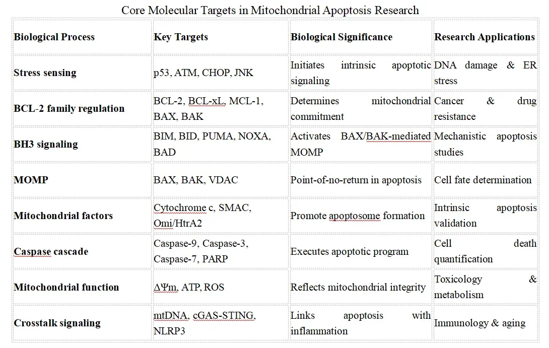 ###Key research trend: Modern mitochondrial apoptosis research is evolving beyond the classical BCL-2–BAX/BAK–cytochrome c–caspase cascade toward an integrated systems biology framework encompassing mitochondrial quality control, organelle dynamics, innate immune signaling, metabolic regulation, and crosstalk with other regulated cell death pathways. Emerging technologies, including single-cell and spatial multi-omics, are enabling mechanistic dissection of apoptosis heterogeneity and supporting precision therapeutic development.
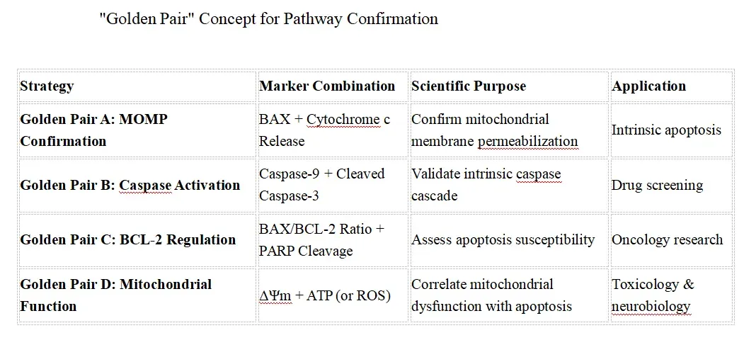
Recommended Mitochondrial Apoptosis Marker Strategy
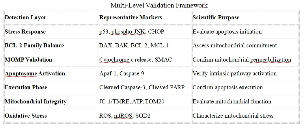
Core Mitochondrial Apoptosis Validation Strategy
Competitive Technology Landscape
Mitochondrial Apoptosis Research Workflow 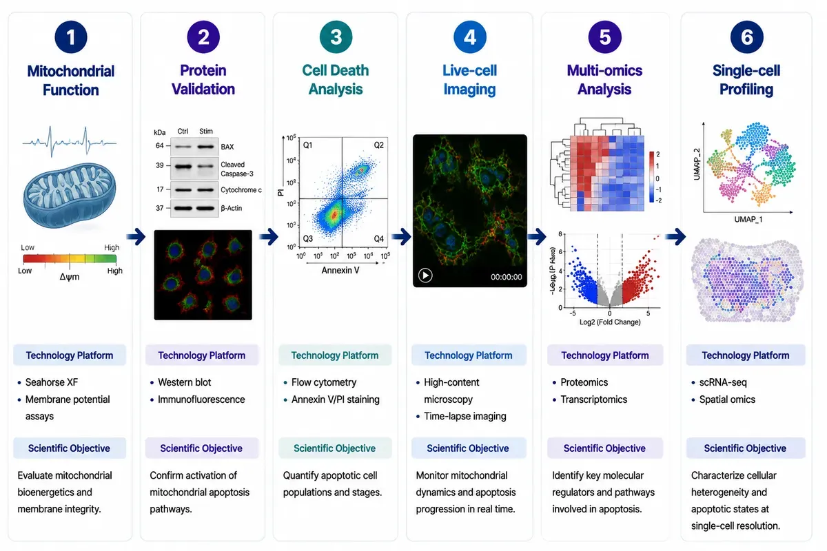 An integrated mitochondrial apoptosis research workflow combining bioenergetic analysis, pathway validation, functional cell death assays, live-cell imaging, multi-omics profiling, and single-cell technologies to elucidate mitochondrial regulation, apoptotic mechanisms, and cellular heterogeneity.
Pathway Overview
Mitochondrial apoptosis is an intrinsic programmed cell death pathway controlled by mitochondrial membrane permeabilization, cytochrome c release, and caspase activation. The BCL-2 family proteins, mitochondrial dynamics, apoptosome formation, and caspase cascade regulate cellular survival and death decisions. This pathway is fundamental to cancer biology, neurodegeneration, immune regulation, and tissue homeostasis. 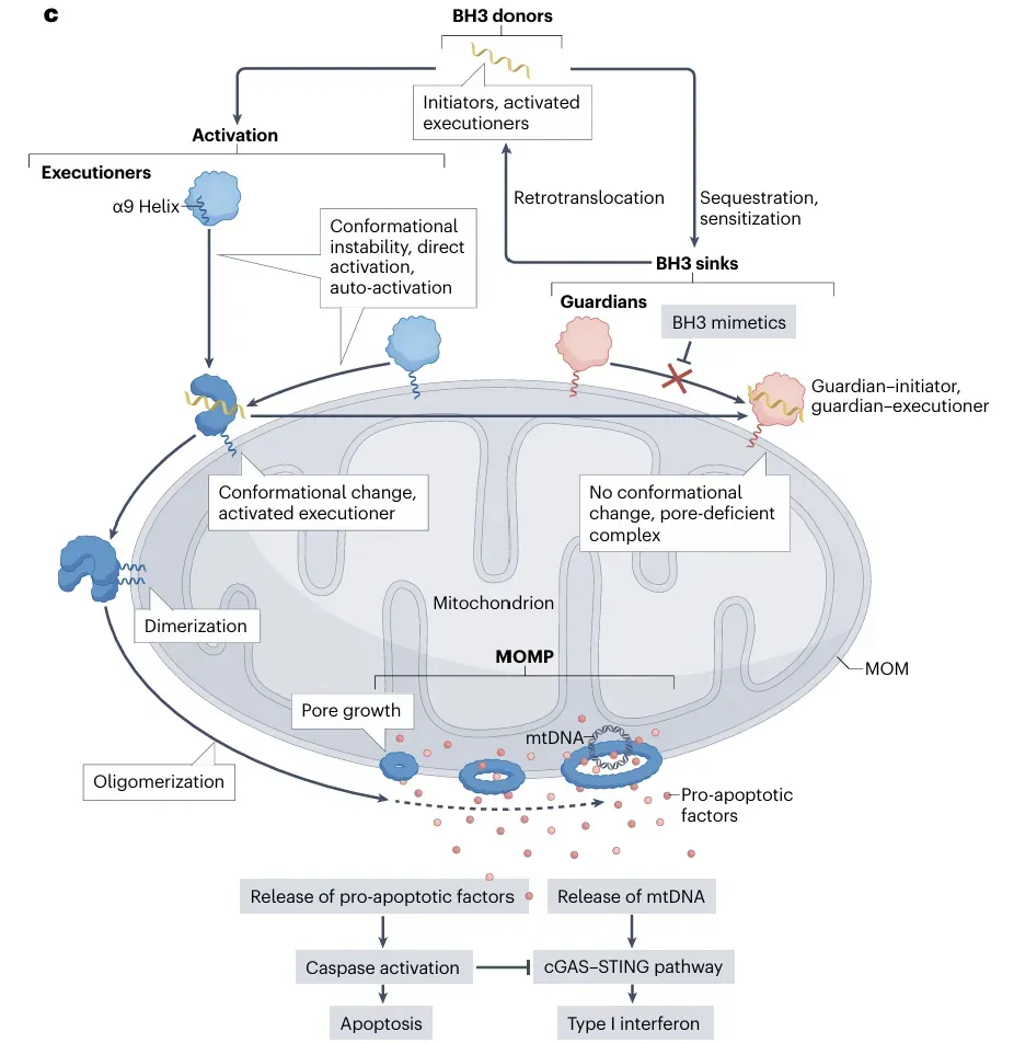 The pivotal event in mitochondria-mediated apoptosis is the rupture of the mitochondrial outer membrane (MOM) following pore formation mediated by executioner members of the BCL-2 family. (PMID: 37438560)
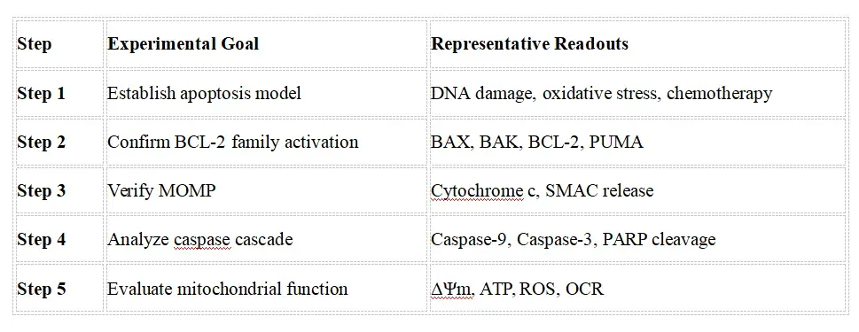
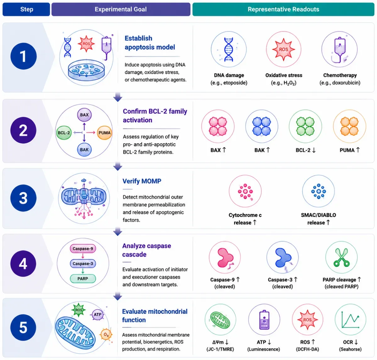
Recommended Experimental Validation Workflow
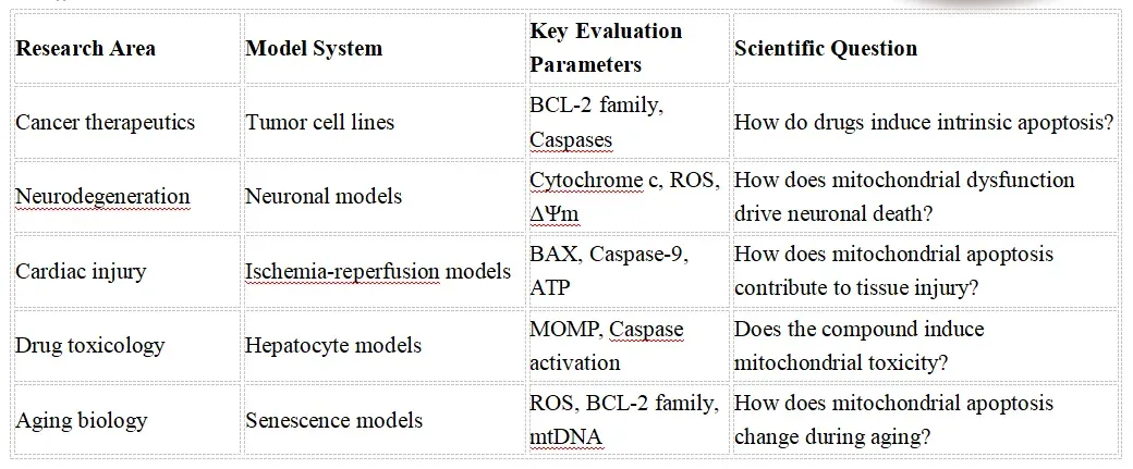
Featured Research Application Examples
Frequently Asked Questions
Q1. Which biomarkers should I use to comprehensively evaluate mitochondrial apoptosis activation? Mitochondrial apoptosis should be assessed using multiple complementary biomarkers across the intrinsic apoptotic pathway. Key markers include BCL-2 family proteins (BAX, BCL-2, BCL-XL, MCL-1) for mitochondrial membrane regulation, cytochrome c release for mitochondrial outer membrane permeabilization (MOMP), cleaved Caspase-9 and cleaved Caspase-3 for apoptotic execution, and PARP cleavage as a hallmark of apoptosis. Combining upstream mitochondrial events with downstream caspase activation provides a comprehensive evaluation of apoptotic signaling. Q2.How can mitochondrial dysfunction be distinguished from mitochondrial apoptosis? Mitochondrial dysfunction does not always lead to apoptosis. Mitochondrial status should be evaluated through bioenergetic and structural measurements, including Seahorse XF analysis, ATP production, oxygen consumption rate (OCR), mitochondrial membrane potential (ΔΨm), and reactive oxygen species (ROS) analysis. Apoptotic commitment requires confirmation of mitochondrial outer membrane permeabilization, cytochrome c release, caspase activation, and cell death execution markers. Q3.Which experimental approaches provide the most reliable assessment of mitochondrial apoptosis? Robust mitochondrial apoptosis studies integrate Western blotting, immunofluorescence, flow cytometry (Annexin V/PI staining), mitochondrial fractionation, caspase activity assays, live-cell imaging, mitochondrial membrane potential assays, and genetic approaches (CRISPR/RNAi). Combining molecular, functional, and imaging-based approaches enables accurate characterization of apoptotic pathway activation and biological consequences. Q4.What are the most common pitfalls in mitochondrial apoptosis research? Common challenges include relying on a single apoptosis marker, confusing general mitochondrial stress with apoptotic commitment, measuring total cytochrome c without assessing mitochondrial release, overlooking the balance between pro- and anti-apoptotic BCL-2 family proteins, and failing to distinguish early apoptotic events from late-stage cell death. Comprehensive analysis requires multiple time points, appropriate controls, and orthogonal validation approaches. Q5.How can mitochondrial apoptosis be comprehensively characterized in physiological and disease contexts? Modern mitochondrial apoptosis research integrates mitochondrial functional analysis, pathway validation, cell death profiling, high-content imaging, multi-omics technologies, and single-cell approaches to define apoptotic regulation, cellular heterogeneity, and disease mechanisms in cancer, neurodegeneration, aging, cardiovascular disorders, and therapeutic response studies.
Key References
1.Glover H.L., Schreiner A., Dewson G., Tait S.W.G. (2024). Mitochondria and cell death. Nature Cell Biology. 26:1434–1446.
2.Czabotar P.E., Garcia-Saez A.J. (2023). Mechanisms of BCL-2 family proteins in mitochondrial apoptosis. Nature Reviews Molecular Cell Biology. 24:732–748.
3.Tábara L.C., Segawa M., Prudent J. (2025). Molecular mechanisms of mitochondrial dynamics. Nature Reviews Molecular Cell Biology. 26:123–146.
4.Yuan J., Ofengeim D. (2024). A guide to cell death pathways. Nature Reviews Molecular Cell Biology. 25:379–395.
5.Vogler M., Braun Y., Smith V.M., et al. (2025). The BCL2 family: from apoptosis mechanisms to new advances in targeted therapy. Signal Transduction and Targeted Therapy.
6.Baker Z.N., Forny P., Pagliarini D.J. (2024). Mitochondrial proteome research: the road ahead. Nature Reviews Molecular Cell Biology. 25:65–82.
7.Saunders T.L., Windley S.P., Gervinskas G., et al. (2024). Exposure of the inner mitochondrial membrane triggers apoptotic mitophagy. Cell Death & Differentiation. 31:335–347.