
info@ucallmlabs.com
Chronic nasal disorders and nasal mucosal infections are highly prevalent upper respiratory diseases worldwide, and their recurrent episodes continuously exacerbate the burden on social healthcare systems. As the primary defensive barrier of the upper respiratory tract, the nasal epithelium represents a highly specialized dynamic mucosal tissue. It integrates physical barrier protection, mucociliary clearance and sophisticated innate immune responses, and directly governs the initiation and progression of various nasal diseases. Dysfunction of the nasal epithelium is not merely a secondary pathological manifestation; instead, it acts as a critical initiating and driving factor for multiple nasal lesions such as chronic rhinosinusitis, allergic rhinitis and viral nasal infections.
At present, basic research on the nasal mucosa mainly relies on animal models and immortalized cell lines. Nevertheless, these systems possess unavoidable inherent limitations, including interspecies biological disparities, aberrant cellular differentiation phenotypes, and an inability to fully reconstruct the three-dimensional tissue architecture of the human nasal mucosa. These drawbacks greatly hinder the mechanistic investigation of nasal diseases and translational research on precise therapeutic strategies.
Nasal organoids can recapitulate the self-assembly characteristics, cellular heterogeneity and disease-specific pathological phenotypes of nasal epithelial tissues within a three-dimensional culture microenvironment. Currently, this model has been adopted for research covering mucosal infection mechanisms, inflammation-induced tissue remodeling and the functional regulation of epithelial ion channels. It provides a reliable in vitro research platform for elucidating the pathogenesis of nasal disorders including chronic rhinosinusitis and advancing individualized precision therapy.
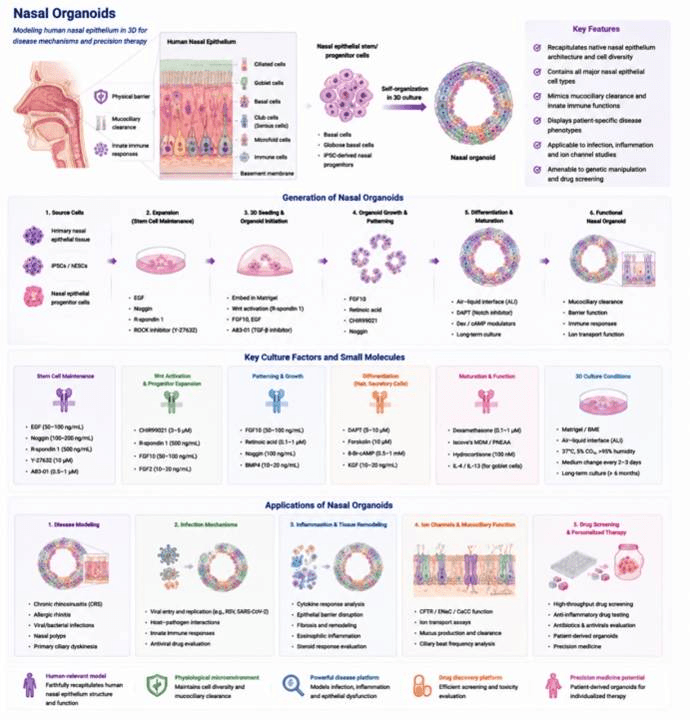
Generation, Culture, and Applications of Nasal Organoids.
Data display.
(A) Organoid culture status (bright-field morphology)
(B) Multicolor immunofluorescence staining
(C) Post-translational modification proteomics
(D) Epigenetic profiling (ChIP-seq / ATAC-seq)
(E) Gene editing validation (CRISPR)