
info@mail.ucallm.com
Neural Development & 3D Neural Models
From 3D Model Establishment to Neural Tissue Maturation and Functional Characterization
A continuum of neural models—from neural stem cells and neural progenitors to 3D neural spheroids and neural organoids—enables researchers to investigate neural development, cellular differentiation, tissue organization, neuronal maturation, neural network formation, disease-associated phenotypes, and translational applications.
How the Nervous System Forms and Functions
From Neural Cells to 3D Neural Systems and Functional Networks
Neural development is a highly coordinated process involving cell fate specification, regional patterning, cell migration, tissue organization, neuronal differentiation, and maturation.
During development, neural stem and progenitor cells progressively acquire specialized cellular identities and organize into increasingly complex neural structures. As neurons mature, neurite extension, synapse formation, and network integration contribute to the establishment of functional neural circuits capable of signal transmission and information processing.
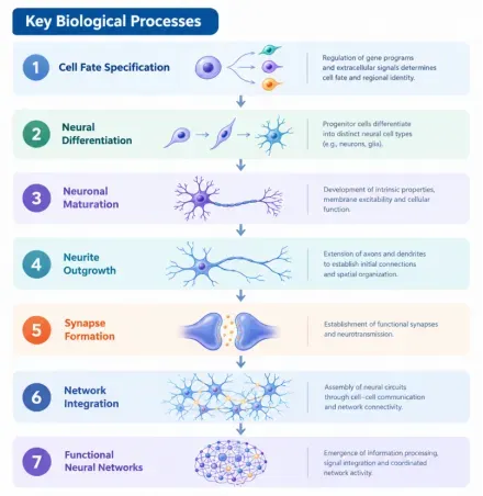
From Neuronal Maturation to Functional Neural Networks — Stepwise progression of neural development toward functional network formation.
A developmental continuum illustrating the transition from immature neural cells to differentiated and mature neurons, followed by neurite extension, synaptic connectivity, network integration, and functional neural activity. From Developmental Mechanisms to Neural Research.
From Developmental Mechanisms to Neural Research
Understanding neural development provides a framework for connecting cellular identity, tissue organization, neuronal maturation, and neural circuit function.
These biological processes support the development and characterization of experimental models for
- Neural Development
- Neuronal Differentiation & Maturation
- Neural Network Formation
- Disease Modeling
- Neurotoxicity & Drug Screening
- Neurodegeneration Research
- Neural Regeneration
By linking developmental biology with advanced model systems, researchers can investigate how molecular and cellular changes contribute to neural dysfunction and disease-associated phenotypes.
Research Tools and Technical Platforms
Neuroscience Cell Culture & Neural Organoid Research Platform — From Neural Cells to 3D Neural Models
An integrated research platform supporting the establishment, maintenance, differentiation, and characterization of neural cell and 3D neural models.
1. Neural Cell Lines
Reproducible Models for Neural Biology & Molecular Mechanisms
Neural cell lines provide accessible and reproducible in vitro systems for investigating neural signaling, gene function, cellular responses, disease-associated mechanisms, and drug effects.
Research Applications
Neural Cell Biology
Cell proliferation · Differentiation · Neurite outgrowth · Cell survival
Molecular Mechanisms
Gene expression · Signaling pathways · Protein function · Cellular responses
Disease Modeling
Disease-associated gene function · Cellular phenotypes · Pathological mechanisms
Drug Research
Compound screening · Mechanism-of-action studies · Neurotoxicity assessment
Molecular Manipulation
Neural cell models can be combined with nucleic-acid delivery and gene-regulation technologies to modulate target expression and investigate molecular mechanisms.
Plasmid DNA · siRNA · miRNA · mRNA · Reporter Systems
Cell Culture → Nucleic-Acid Delivery → Gene Regulation → Molecular Readout
From stable neural cell models to molecular mechanism studies, UcallM provides essential tools for neural-cell culture, transfection, and functional characterization.
Supporting Products
Serum for Reliable Cell Culture & Neural Model Establishment
| Item # | Product Name | Specification | Application |
|---|---|---|---|
| C2740 | Fetal Bovine Serum (Superfine) | 50mL×10 | Various tumor cells and common cell lines |
| C2741 | Fetal Bovine Serum (Superfine) | 500mL | Various tumor cells and common cell lines |
Data display:
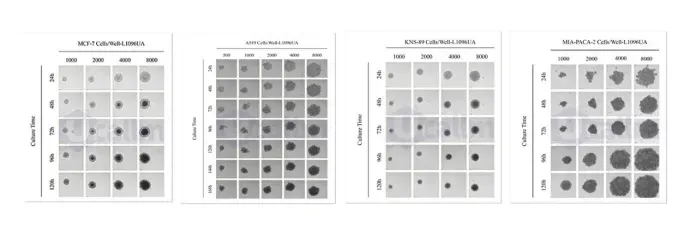
MCF-7, A549, KNS-89, and MIA PaCa-2 tumor cell lines form 3D tumor spheroids when cultured in Ucallm® Ultra-Low Attachment U-Bottom Plates (Cat. No. L1096UA).
Transfection Reagents
Efficient Nucleic Acid Delivery for Cell-Based Neuroscience Research
| Product Name | Cat. No. | Reactivity |
|---|---|---|
| PerFect | TF01 | Broad Compatibility with Diverse Tumor Cells and Cell Lines. |
| LipoTrans | TF06 | Broad Compatibility with Mammalian Cell Lines, including suspension cell types. |
| RNAMute | TF08 | Suitable for various Mammalian Cell Lines. |
| PerMute | TF09 | Suitable for various Mammalian Cell Lines. |
| BL-LNP-MC3-200 | TF12 | Broad Compatibility with Mammalian Cell Lines |
| BL-LNP-ALC-200 | TF13 | Broad Compatibility with Mammalian Cell Lines |
| BL-LNP-SM102-200 | TF14 | Broad Compatibility with Mammalian Cell Lines |
2. Primary Neural Cells
Physiologically Relevant Models for Neural Cell Function
Primary neural cells are isolated directly from neural tissues, including the brain, spinal cord, and peripheral nervous system. Compared with continuously passaged cell lines, primary cells can better preserve tissue-associated morphology, molecular characteristics, and cellular functions.
Representative Neural Cell Types
Primary Neurons
Neurite outgrowth · Axon and dendrite development · Synaptogenesis · Neural network formation
Primary Astrocytes
Neuronal support · Metabolic regulation · Reactive responses · Microenvironment remodeling
Primary Oligodendrocytes
Lineage differentiation · Myelination · Axonal support · White matter biology
Primary Microglia
Neuroimmune signaling · Inflammatory responses · Phagocytosis · Neural injury responses


🧠 Workflow and main features of primary neural cell culture
Tissue Isolation
Enzymatic / Mechanical DissociationCell SeedingCulture & MaturationPhenotypic CharacterizationFunctional / Molecular Analysis
Molecular Manipulation
Primary neural cells can be combined with nucleic-acid delivery technologies for:
Gene Overexpression · Gene Silencing · Reporter Assays · Functional Validation · Signaling Studies
We support primary neural-cell workflows from culture and maturation to nucleic-acid delivery, phenotypic profiling, and molecular mechanism analysis.
Supporting Products
Serum – Reliable Culture Support for Cell-Based Research
| Item # | Product Name | Specification | Application |
|---|---|---|---|
| C275 | Fetal Bovine Serum (Premium) | 50 mL × 10 | Common cell lines, primary cells, stem cells, immune cells, etc. |
| C275 | Fetal Bovine Serum (Premium) | 500 mL | Common cell lines, primary cells, stem cells, immune cells, etc. |
Transfection Reagents – Efficient Nucleic Acid Delivery Across Diverse Cell Models
| Product Name | Cat. No. | Reactivity |
|---|---|---|
| PolyPer | TF02 | Broad Compatibility with Mammalian Cell Lines, Primary Cells, Hard-to-Transfect Cells, Insect Cell Lines. |
| PolyPer plus | TF03 | Suitable for common Mammalian Cell Lines, Primary Cells, and Hard-to-Transfect Cells. |
| PolyPer max | TF04 | Suitable for most cell types, including Primary Cells, Hard-to-Transfect Cells, and Toxicity-Sensitive Cells, with efficient DNA delivery into both Mammalian Cell Lines and Insect Cells. |
| LipoEffect | TF05 | Suitable for HEK293 and CHO Cells. Effective in Primary Cells and Hard-to-Transfect Cells. |
| LongTrans | TF07 | Suitable for various Mammalian Cell Lines, including Hard-to-Transfect Mammalian Cells. |
| PowerTrans | TF10 | Broad Compatibility with Mammalian Cell Lines and Hard-to-Transfect Cells. |
| RNAMute plus | TF11 | Broad Compatibility with Mammalian Cell Lines and Hard-to-Transfect Cells. |
3. Neural Stem Cells
Modeling Neural Development, Differentiation & Cell Fate
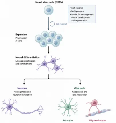
Neural stem cells (NSCs) are self-renewing, multipotent neural progenitors capable of generating major neural lineages, including neurons, astrocytes, and oligodendrocytes under appropriate culture conditions.
Key Research Areas
Neural Development
Study neurogenesis, lineage specification, and developmental progression.
Cell-Fate Determination
Investigate molecular mechanisms controlling neuronal and glial differentiation.
Neural Regeneration
Explore neural progenitor responses, differentiation, and regenerative potential.
Disease Modeling
Generate controlled cellular models for neurological disease mechanisms.
Drug & Neurotoxicity Research
Evaluate compound effects on neural differentiation, survival, and maturation.
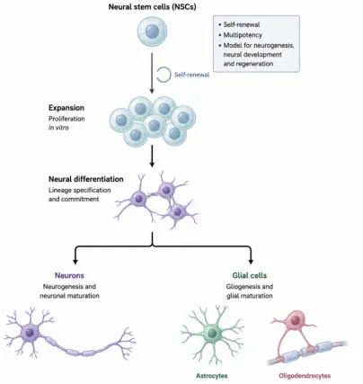
🧬 Multipotent properties and application scenarios of neural stem cells.
Model Integration
NSCs can serve as a flexible starting point for:
2D Neural Cultures · 3D Neural Spheroids · Neural Organoids
UcallM Research Support
NSC Expansion → Differentiation → Lineage Characterization → Functional Profiling
By connecting stem-cell biology with controlled differentiation and molecular characterization, we support investigation of neural development, cell fate, disease mechanisms, and regenerative biology.
Recommended Products
Neural Signaling Proteins for Cell Differentiation, Maturation & 3D Model Development
| SKU | Molecule | Species | Source | Tags | Accession | Protein Length |
|---|---|---|---|---|---|---|
| PR00044 | BDNF | Human/Mouse/Rat | E. coli | Tag free | P23560 | His129-Arg247 |
| PR00326 | BMP4 | Human | E. coli | Tag free | P12644 | Ser293-Arg408 |
| PR00055 | CNTF | Human | E. coli | Tag free | P26441 | Ala2-Met200 |
| PR00016 | EGF | Human | E. coli | Tag free | P01133 | Asn971-Arg1023 |
| PR00167 | EGF | Mouse | E. coli | C-6His | P01132 | Asn977-Arg1029 |
| PR00244 | FGF10 | Human | E. coli | Tag free | O15520 | Gln38-Ser208 |
| PR00344 | FGF10 | Mouse | E. coli | Tag free | O35565 | Ser62-Thr209 |
| PR00025 | FGF2/bFGF | Mouse | E. coli | Tag free | P15655 | Met1-Ser154 |
| PR00027 | FGF2/bFGF | Human | E. coli | Tag free | P09038-4 | Gly132-Ser288 |
| PR00094 | FGF2/bFGF | Human | E. coli | Tag free | P09038-4 | Met134-Ser288 |
| PR00FGF2/ | Human | E. coli | Tag | P09038-4 | Pro143-Ser288 |
| SKU | Molecule | Species | Source | Tags | Accession | Protein Length |
|---|---|---|---|---|---|---|
| PRO0249 | PDGF-BB | Mouse | E. coli | C-6His | AAH53430.1 | Ser82-Thr190 |
| PRO0050 | SHH | Human | E. coli | Tag free | Q15465 | Cys24-Gly197 (C24II) |
| PRO0057 | SHH | Human | E. coli | Tag free | Q15465 | Cys24-Gly197 |
| PRO0173 | SHH | Mouse | E. coli | Tag free | Q62226 | Cys25-Gly198 (C25II) |
| PRO0175 | SHH | Mouse | E. coli | Tag free | Q62226 | Cys25-Gly198 |
| PRO0287 | SHH | Human | HEK293 | C-6His | Q15465 | Cys24-Gly197 |
| PRO0290 | SHH | Mouse | HEK293 | C-6His | Q62226 | Cys25-Gly198 |
| PRO0375 | VEGFA/VEGF165 | Human | HEK293 | Tag free | P15692-4 | Ala27-Arg191 |
| PRO2601 | VEGFA/VEGF165 | human | HEK293 | C-6His-avi | P15692-4 | Ala27-Arg191 |
| PRO2756 | VEGFA/VEGF165 | Human | HEK293 | N-6His | P15692-4 | Ala27-Arg191 |
| PR16890 | VEGFA/VEGF165 | Human | E. coli | C-6His | NP_001165097 | Ala27-Arg191 |
| PR16984 | VEGFA/VEGF165 | Mouse | E. coli | C-6His | NP_033531.3 | Ala205-Arg368 |
High-Quality Proteins for Reliable Research
We provide recombinant proteins and cytokines designed for high biological performance, consistent quality, and reproducible experimental outcomes.
Five Core Advantages
High Purity & Biological Activity
High-quality proteins with validated biological activity.
Low Endotoxin
Controlled endotoxin levels to support sensitive cellular and organoid applications.
Batch-to-Batch Consistency
Rigorous quality management ensures consistent product performance across batches.
Comprehensive Portfolio
A broad range of recombinant proteins and cytokines supporting diverse research applications.
Stable & Reliable Performance
Validated performance across multiple experimental platforms.
Serum support for a broad range of cell-based research
| Item # | Product Name | Specification | Application |
|---|---|---|---|
| C2770 | Fetal Bovine Serum (Superfine), Origin: Uruguay | 50 mL × 10 | Common cell lines, most primary cells, stem cells, immune cells, etc. |
| C2771 | Fetal Bovine Serum (Superfine), Origin: Uruguay | 500 mL | Common cell lines, most primary cells, stem cells, immune cells, etc. |
4. 3D Neural Spheroids
Bridging 2D Cell Culture and Complex Neural Tissues
3D neural spheroids are multicellular three-dimensional models generated through cell aggregation and self-organization under low-attachment or non-adherent culture conditions.
Compared with conventional 2D cultures, 3D spheroids provide a tissue-like spatial environment that enables more complex investigation of cellular interactions, extracellular matrix relationships, and local microenvironmental changes.
🧫 Schematic diagram of 3D neural spheroid generation and key features.
Key Features
3D Architecture
Spatial organization beyond planar cell culture
Enhanced Cell-Cell Interaction
More complex cellular communication within a three-dimensional environment
Cell-ECM Interactions
Investigation of extracellular matrix and microenvironmental effects
Differentiation & Maturation
Support neural differentiation, neurite development, and network formation
3D neural spheroids provide a practical intermediate model for investigating neural biology in a more physiologically relevant three-dimensional context.
Data presentation: Neural Cells cultured with UcallM® Ultra-Low Attachment Surface Culture Plates
| Cell | Cell Type | Main Neuroscience Applications |
|---|---|---|
| KNS-89 | Human glioma cell line | Brain tumor research, glioma stemness, tumor cell proliferation, drug screening, tumor invasion and drug resistance studies |
| mBM-MSCs | Mouse bone marrow mesenchymal stem cells | Neural regeneration, cell therapy, tissue repair, cellular paracrine effects, neuroprotection research |
| mHN / mHNCs | Mouse hippocampal neurons / neural cells | Neuronal survival, neurite outgrowth, neurotoxicity, neuroprotection, neurodegenerative disease models |
| Mouse spinal cord neurons | Mouse spinal cord neurons | Spinal cord injury, neural regeneration, neural repair, neuroprotection, neurotoxicity research |
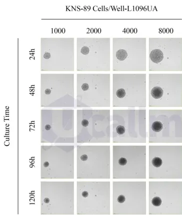
📈 KNS-89 cells cultured on UcallM® Ultra-Low Attachment Surface forms tumor spheroids. KNS-89 cells were planted in 96-well tissue culture plates and ultra-low attachment plates at concentrations of 500, 1000, 2000, 4000, and 8000 cells per well. Live-cell imaging was conducted at 24, 48, 72, 96, and 120 hours after seeding. Scale bars represent 200 μm.
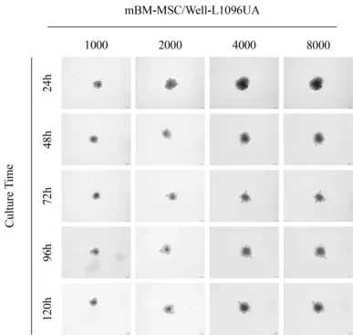
📈 mBM-MSCs cultured on UcallM® Ultra-Low Attachment Surface forms tumor spheroids. mBM-MSCs were planted in 96-well tissue culture plates and ultra-low attachment plates at concentrations of 500, 1000, 2000, 4000, and 8000 cells per well. Live-cell imaging was conducted at 24, 48, 72, 96, and 120 hours after seeding. Scale bars represent 200 μm.
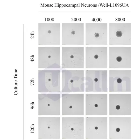
📈 mHN cultured on UcallM® Ultra-Low Attachment Surface forms tumor spheroids. mHNCs were planted in 96-well tissue culture plates and ultra-low attachment plates at concentrations of 500, 1000, 2000, 4000, and 8000 cells per well. Live-cell imaging was conducted at 24, 48, 72, 96, and 120 hours after seeding. Scale bars represent 200 μm.
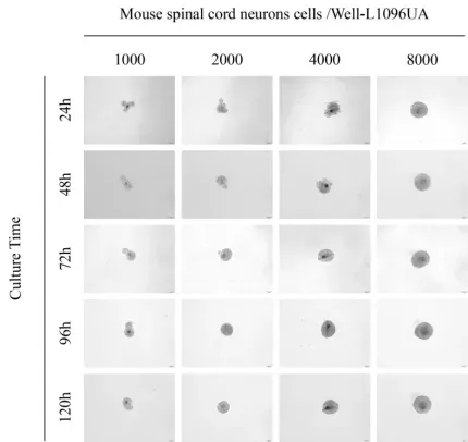
📈 Mouse spinal cord neurons cultured on UcallM® Ultra-Low Attachment Surface forms tumor spheroids. Mouse spinal cord neurons were planted in 96-well tissue culture plates and ultra-low attachment plates at concentrations of 500, 1000, 2000, 4000, and 8000 cells per well. Imaging was conducted at 24, 48, 72, 96, and 120 hours after seeding. Scale bars represent 200 μm.
5. Neural Organoids
From Cells to Tissue-Like Neural Models
Neural organoids are three-dimensional neural tissue models generated from pluripotent stem cells or other stem-cell sources through 3D culture and directed differentiation.
They can reproduce selected aspects of neural development, cellular composition, tissue organization, and cell-cell interactions, providing human-relevant experimental systems for studying complex neural biology.
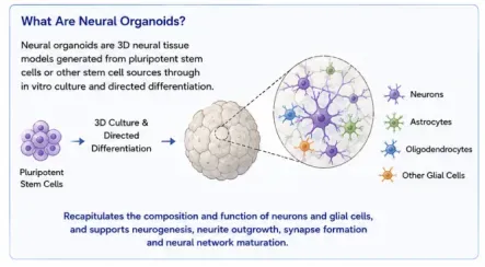
Schematic illustration of neural organoid generation and cellular composition.
Key Biological Features
Cellular Diversity
Neurons · Astrocytes · Oligodendrocytes · Other neural and glial populations
Spatial Organization
Three-dimensional tissue architecture and cellular organization
Neural Development
Neurogenesis · Differentiation · Neurite outgrowth · Maturation
Cell-Cell Interaction
Neuronal-glial interactions · Synaptogenesis · Neural microenvironment
Functional Maturation
Neural network formation · Activity-associated responses
Research Applications
Neural Development
Model human neural development and cellular differentiation.
Neurodegeneration
Investigate disease-associated molecular and cellular phenotypes.
Neuroinflammation
Study interactions between neural cells and immune-related components.
Neural Tumor Research
Investigate tumor-associated neural microenvironments and cellular phenotypes.
Drug Screening & Neurotoxicity
Evaluate compound responses in human-relevant 3D neural models.
Translational Research
Support mechanism studies and preclinical research using advanced human-relevant models.
From Animal Models to Human-Relevant Systems — Regulatory momentum is accelerating the transition toward standardized, reproducible 3D models, organoids, and organ-on-a-chip technologies for next-generation neuroscience research.
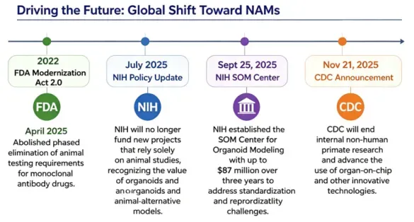
Driving the future: global shift toward New Alternative Methods (NAMs).
Neural Organoid Solutions
From Cells to Tissues - Building Standardized, Human-Relevant 3D Models
We provide standardized and reproducible organoid solutions integrating 3D culture, vascularization, and multi-cellular co-culture technologies to support human-relevant disease modeling, drug screening, pharmacodynamic evaluation, and mechanistic research.
Core Capabilities
Standardized Organoid Generation
Established culture systems and technical platforms support the efficient generation of diverse organoid models, with a reported model generation success rate of >95%.
Vascularization
Vascularization strategies promote the development of vascular networks and tissue architecture, supporting oxygen and nutrient supply, long-term tissue maintenance, and functional maturation.
Multi-cellular Co-culture
Integration of neurons, glial cells, immune cells, fibroblasts, and endothelial cells enables investigation of cell-cell communication and microenvironmental interactions within complex 3D tissues.
Enhanced Physiological Relevance
Advanced organoid models can better recapitulate key aspects of native tissues, including cellular composition, spatial organization, tissue architecture, and biological interactions.
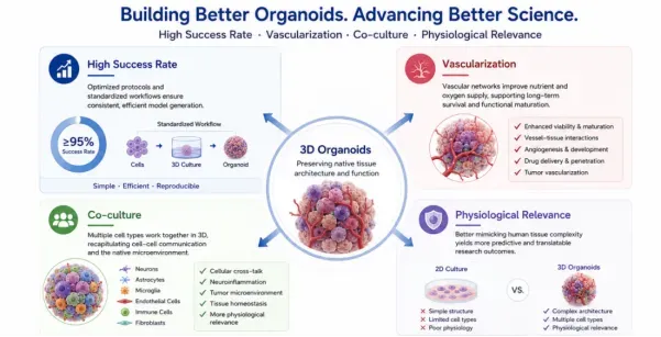
Key advantages and broad applications of advanced 3D organoid models.
Advanced 3D Organoids Models
From Cellular Models to Human-Relevant Tissue Systems
| 01 | 02 | 03 | 04 |
|---|---|---|---|
| Standardized Generation Reproducible culture & differentiation | Vascularization Long-term maturation & neurovascular interactions | Multicellular Culture Neural & non-neural cell interactions | Physiological Relevance 3D architecture & cellular heterogeneity |
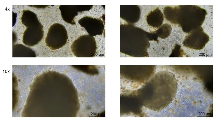
ID: BR144-D6
Species: Human
Sample Type: Human brain organoid
Time Point: P0 Day 6
Basement Membrane Matrix
Designed to provide a supportive extracellular matrix environment for organoid growth, expansion, differentiation, and 3D morphogenesis.
| Cat. No. | Product Name | Pack Size |
|---|---|---|
| C2800 | Ucell® Matrix Standard | 2 mL |
| C2801 | Ucell® Matrix Standard | 5 mL |
| C2802 | Ucell® Matrix Standard | 10 mL |
| C2810 | Ucell® Matrix Standard, Phenol Red-Free | 2 mL |
| C2811 | Ucell® Matrix Standard, Phenol Red-Free | 5 mL |
| C2812 | Ucell® Matrix Standard, Phenol Red-Free | 10 mL |
| C2820 | Ucell® Matrix Reduced Growth Factor | 2 mL |
| C2821 | Ucell® Matrix Reduced Growth Factor | 5 mL |
| C2822 | Ucell® Matrix Reduced Growth Factor | 10 mL |
| C2830 | Ucell® Matrix Reduced Growth Factor, Phenol Red-Free | 2 mL |
| C2831 | Ucell® Matrix Reduced Growth Factor, Phenol Red-Free | 5 mL |
| C2832 | Ucell® Matrix Reduced Growth Factor, Phenol Red-Free | 10 mL |
| C2860 | Ucell® Matrix High Concentration, Standard | 2 mL |
| C2861 | Ucell® Matrix High Concentration, Standard | 5 mL |
| C2862 | Ucell® Matrix High Concentration, Standard | 10 mL |
| C2870 | Ucell® Matrix High Concentration, Standard, Phenol Red-Free | 2 mL |
| C2871 | Ucell® Matrix High Concentration, Standard, Phenol Red-Free | 5 mL |
| C2872 | Ucell® Matrix High Concentration, Standard, Phenol Red-Free | 10 mL |
| C2880 | Ucell® Matrix High Concentration, Reduced Growth Factor | 2 mL |
| C2881 | Ucell® Matrix High Concentration, Reduced Growth Factor | 5 mL |
| C2882 | Ucell® Matrix High Concentration, Reduced Growth Factor | 10 mL |
| C2840 | Ucell® Matrix for Organoid Culture | 2 mL |
| C2841 | Ucell® Matrix for Organoid Culture | 5 mL |
| C2842 | Ucell® Matrix for Organoid Culture | 10 mL |
| C2850 | Ucell® Matrix hESC-Qualified Matrigel, for Human Embryonic Stem Cell Culture | 2 mL |
| C2851 | Ucell® Matrix hESC-Qualified Matrigel, for Human Embryonic Stem Cell Culture | 5 mL |
| C2852 | Ucell® Matrix hESC-Qualified Matrigel, for Human Embryonic Stem Cell Culture | 10 mL |
🔬 Gastric cancer (GC) tumor-derived organoids.


Bright-field and fluorescence imaging of gastric cancer-derived organoids cultured using Pharmadea Specialty Basement Membrane Matrix (C2842).
Recombinant Proteins
Key Growth Factors & Signaling Molecules for Neural Development and 3D Neural Models
Support stem-cell maintenance, neural lineage specification, tissue patterning, maturation, vascularization, and microenvironment modeling across advanced neural culture and organoid workflows.
| SKU | Molecule | Species | Source | Tags | Accession | Protein Length |
|---|---|---|---|---|---|---|
| PR00087 | Activin A/INHBA | Human/Mouse/Rat | HEK293 | Tag free | P08476 | Gly311-Ser426 |
| PR00044 | BDNF | Human/Mouse/Rat | E. coli | Tag free | P23560 | His129-Arg247 |
| PR00007 | BMP2 | Human/Mouse/Rat | E. coli | Tag free | P12643 | Gln283-Arg396 |
| PR00326 | BMP4 | Human | E. coli | Tag free | P12644 | Ser293-Arg408 |
| PR00327 | BMP7 | Human | HEK293 | Tag free | P18075 | Ser293-His431 |
| PR00055 | CNTF | Human | E. coli | Tag free | P26441 | Ala2-Met200 |
| PR00059 | CXCL12/SDF-1 | Human | E. coli | Tag free | P48061 | Ser19-Met93 |
| PR00060 | CXCL12/SDF-1 | Human | E. coli | Tag free | P48061 | Lys22-Lys89 |
| PR00061 | CXCL12/SDF-1 | Human | E. coli | Tag free | P48061 | Lys22-Met93 |
| PR00091 | CXCL12/SDF-1 | Mouse | E. coli | Tag free | P40224 | Lys22-Lys89 |
| PR00676 | DKK1 | Human | HEK293 | N-8His | O94907 | Thr32-His266 |
| SKU | Molecule | Species | Source | Tags | Accession | Protein Length |
|---|---|---|---|---|---|---|
| PR02515 | DKK1 | Human | HEK293 | C-6His | O94907 | Thr32-His266 |
| PR00016 | EGF | Human | E. coli | Tag free | P01133 | Asn971-Arg1023 |
| PR00167 | EGF | Mouse | E. coli | C-6His | P01132 | Asn977-Arg1029 |
| PR00244 | FGF10 | Human | E. coli | Tag free | O15520 | Gln38-Ser208 |
| PR00344 | FGF10 | Mouse | E. coli | Tag free | O35565 | Ser62-Thr209 |
| PR00159 | FGF19 | Human | E. coli | N-6His | O95750 | Phe27-Lys216 |
| PR00025 | FGF2/bFGF | Mouse | E. coli | Tag free | P15655 | Met1-Ser154 |
| PR00027 | FGF2/bFGF | Human | E. coli | Tag free | P09038-4 | Gly132-Ser288 |
| PR00094 | FGF2/bFGF | Human | E. coli | Tag free | P09038-4 | Met134-Ser288 |
| PR00098 | FGF2/bFGF | Human | E. coli | Tag free | P09038-4 | Pro143-Ser288 |
| PR00180 | FGF2/bFGF | Human | E. coli | Tag free | BAG70135.1 | Met1-Ser155 |
| PR00222 | FGF2/bFGF | Rat | E. coli | Tag free | P13109 | Ala11-Ser154 |
| PR00354 | FGF2/bFGF | Human | E. coli | Tag free | BAG70264.1 | Met1-Ser155 |
| PR00242 | FGF4 | Human | E. coli | Tag free | P08620 | Ser54-Leu206 |
| PR00285 | FGF4 | Mouse | E. coli | Tag free | P11403 | Ser67Leu202 |
| PR00286 | FGF4 | Human | E. coli | Tag free | P08620 | Ser71-Leu206 |
| PR00101 | FGF8 | Human/Mouse | E. coli | Tag free | P55075-3/P37237-2 | Gln23-Arg215 |
| PR00013 | IGF1 | Human | E. coli | Tag free | P05019 | Gly49-Ala118 |
| PR00017 | IGF1 | Human | E. coli | Tag free | P05019 | Thr52-Ala118 |
| PR00018 | IGF1 | Human | E. coli | Tag free | P05019 | Gly49-Ala118 |
| PR00248 | IGF1 | Mouse | E. coli | C-6His | P05017 | Gly49-Ala118 |
| SKU | Molecule | Species | Source | Tags | Accession | Protein Length |
|---|---|---|---|---|---|---|
| PR00343 | IL6 | Mouse | HEK293 | N-6His | P08505 | Phe25-Thr211 |
| PR00361 | IL6 | Human | E. coli | Tag free | P05231 | Val30-Met212 |
| PR01002 | IL6 | Mouse | E. coli | Tag free | P08505 | Phe25-Thr211 |
| PR02813 | IL6 | Rhesus Macaque | HEK293 | C-6His | A0A1D5QRZ4 | Ala28-Met212 |
| PR00360 | Insulin | Human | P. pastoris | Tag free | Confidential | Confidential |
| PR00015 | NOG/Noggin | Mouse | HEK293 | C-6His | P97466 | Gln28-Cys232 |
| PR00122 | NOG/Noggin | Human | HEK293 | Tag free | Q13253 | Gln28-Cys232 |
| PR00047 | NTF3 | Human | E. coli | Tag free | P20783 | Tyr139-Thr257 |
| PR00065 | PDGF-BB | Human | E. coli | Tag free | P01127 | Ser82-Thr190 |
| PR00249 | PDGF-BB | Mouse | E. coli | C-6His | AAH53430.1 | Ser82-Thr190 |
| PR00275 | RSPO1 | Human | HEK293 | C-6His | Q2MKA7 | Ser21-Ala263 |
| PR00336 | RSPO1 | Mouse | HEK293 | C-Flag | NP_619624.2 | Ser21-Gln265 |
| PR00020 | SCF/KITLG | Human | E. coli | Tag free | P21583 | Glu26-Ala189 |
| PR00097 | SCF/KITLG | Mouse | E. coli | Tag free | P20826 | Lys26-Ala189 |
| PR00119 | SCF/KITLG | Mouse | HEK293 | C-6His | P20826 | Lys26-Ala189 |
| PR00120 | SCF/KITLG | Rat | HEK293 | C-6His | P21581 | Gln26-Ala189 |
| PR00155 | SCF/KITLG | Rat | E. coli | C-6His | P21581 | Gln26-Ala189 |
| PR00271 | SCF/KITLG | Human | HEK293 | C-6His | P21583 | Glu26-His214 |
| PR00050 | SHH | Human | E. coli | Tag free | Q15465 | Cys24-Gly197 (C24II) |
| PR00057 | SHH | Human | E. coli | Tag free | Q15465 | Cys24-Gly197 |
| PR00173 | SHH | Mouse | E. coli | Tag free | Q62226 | Cys25-Gly198 (C25II) |
| SKU | Molecule | Species | Source | Tags | Accession | Protein Length |
|---|---|---|---|---|---|---|
| PR00175 | SHH | Mouse | E. coli | Tag free | Q62226 | Cys25-Gly198 |
| PR00287 | SHH | Human | HEK293 | C-6His | Q15465 | Cys24-Gly197 |
| PR00290 | SHH | Mouse | HEK293 | C-6His | Q62226 | Cys25-Gly198 |
| PR00110 | TGFB1/TGF-beta-1/LAP | Human | CHO | Tag free | P01137 | Ala279-Ser390 |
| PR00111 | TGFB1/TGF-beta-1/LAP | Human | HEK293 | N-6His | P01137 | Leu30-Ser390(C33S) |
| PR00208 | TGFB1/TGF-beta-1/LAP | Mouse/Rat | HEK293 | Tag free | P04202 | Ala279-Ser390 |
| PR00297 | TGFB1/TGF-beta-1/LAP | Human | HEK293 | N-Avi | P01137 | Ala279-Ser390 |
| PR01781 | TGFB1/TGF-beta-1/LAP | Human | HEK293 | Tag free | P01137 | Leu30-Arg278(C33S) |
| PR02492 | TGFB1/TGF-beta-1/LAP | Mouse | HEK293 | N-8His | P04202 | Leu30-Ser390 (C33S) |
| PR02493 | TGFB1/TGF-beta-1/LAP | Rhesus Macaque | HEK293 | N-6His | F7HCV5 | Leu30-Ser390 (C33S) |
| PR02494 | TGFB1/TGF-beta-1/LAP | Mouse | HEK293 | N-8His-Avi | P04202 | Leu30-Ser390 (C33S) |
| PR02497 | TGFB1/TGF-beta-1/LAP | Human | HEK293 | N-6His-Avi | P01137 | Leu30-Arg278 (C33S) |
| PR00375 | VEGFA/VEGF165 | Human | HEK293 | Tag free | P15692-4 | Ala27-Arg191 |
| PR02601 | VEGFA/VEGF165 | human | HEK293 | C-6His-avi | P15692-4 | Ala27-Arg191 |
| PR02756 | VEGFA/VEGF165 | Human | HEK293 | N-6His | P15692-4 | Ala27-Arg191 |
| PR16890 | VEGFA/VEGF165 | Human | E. coli | C-6His | NP_001165097 | Ala27-Arg191 |
| PR16984 | VEGFA/VEGF165 | Mouse | E. coli | C-6His | NP_033531.3 | Ala205-Arg368 |
| PR00319 | WNT3A | Human | HEK293 | N-fusion | P56704 | Ser19-Lys352 |
Stem Cell Maintenance → Differentiation → Patterning → Maturation → Vascularization → Microenvironment Modeling → Translational Research
1. Imaging, Cell Analysis & Fluorescent Probes
Visualize Cellular Phenotypes and Functional Changes in Neural Models
A comprehensive portfolio of fluorescent probes and cell-analysis reagents supports the visualization and functional characterization of neural cells, 3D neural models, and organoids—from proliferation and viability to apoptosis, mitochondrial function, cytoskeletal remodeling, organelle dynamics, and cellular morphology.
| Research Area | Neuroscience Application | Specific Product | Primary Use | Cat. No. |
|---|---|---|---|---|
| Cell Proliferation | Neural stem/progenitor or cell proliferation, glial cell proliferation, cell proliferation after neural injury | Universal BF™ 488 Click-iT EdU Cell Proliferation Assay Kit | DNA synthesis and cell proliferation detection | C20501 |
| Cell Proliferation | Neural stem/progenitor or cell proliferation, glial cell proliferation, cell proliferation after neural injury | Universal BF™ 555 Click-iT EdU Cell Proliferation Assay Kit | DNA synthesis and cell proliferation detection | C20502 |
| Cell Proliferation | Neural stem/progenitor or cell proliferation, glial cell proliferation, cell proliferation after neural injury | Universal BF™ 594 Click-iT EdU Cell Proliferation Assay Kit | DNA synthesis and cell proliferation detection | C20503 |
| Research Area | Neuroscience Application | Specific Product | Primary Use | Cat. No. |
|---|---|---|---|---|
| Cell Proliferation | Neural stem/progenitor cell proliferation, glial cell proliferation, cell proliferation after neural injury | Universal BF™ 647A Click-iT EdU Cell Proliferation Assay Kit | DNA synthesis and cell proliferation detection | C20505 |
| Cell Viability/Cytotoxicity | Neurotoxicity, neuroprotection, neurodegenerative diseases, drug screening | Universal Live/Dead Cell Viability Assay Kit (SYTO LiveTM 9, PI) | Live/dead cell detection | C20604 |
| Cell Viability/Cytotoxicity | Neurotoxicity, neuroprotection, neurodegenerative diseases, drug screening | Animal Live/Dead Cell Viability/Cytotoxicity Assay Kit | Cell viability, cytotoxicity, and nuclear staining | C20705 |
| Cell Senescence | Neuronal senescence, neurodegenerative diseases, chronic stress and neural injury | β-Galactosidase Cell Senescence Staining Kit | Cell senescence detection | C21001 |
| Research Area | Neuroscience Application | Specific Product | Primary Use | Cat. No. |
|---|---|---|---|---|
| Cell Cycle | Neurogenesis, glial cell proliferation, glioma research | PI Cell Cycle Assay Kit | DNA content, cell cycle, and apoptosis analysis | C21101 |
| Apoptosis | Neuronal injury, neurodegenerative diseases, neurotoxicity, neuroprotection, brain/spinal cord injury | PanSpatio™ Live-Cell Caspase-3/7 Activity and Annexin V Dual Apoptosis Detection Kit | Caspase-3/7 and PS externalization detection | C21204 |
| Apoptosis | Neuronal injury, neurodegenerative diseases, neurotoxicity, neuroprotection, brain/spinal cord injury | PanSpatio™ Live-Cell Caspase-3/7 Activity and Annexin V Dual Apoptosis Detection Kit (Enhanced) | Multiparameter apoptosis detection | C21206 |
| Apoptosis | Neuronal injury, neurodegenerative diseases, neurotoxicity, neuroprotection, brain/spinal cord injury | BF™ 594-Annexin V / Hoechst 33342 Apoptosis Kit | Discrimination and quantitation of live and apoptotic cells | C21407 |
| Apoptosis | Neuronal injury, neurodegenerative diseases, neurotoxicity, neuroprotection, brain/spinal cord injury | One-Step Universal BF™ 488 TUNEL Apoptosis Kit | DNA fragmentation detection | C21501 |
| Research Area | Neuroscience Application | Specific Product | Primary Use | Cat. No. |
|---|---|---|---|---|
| Apoptosis | Neuronal injury, neurodegenerative diseases, neurotoxicity, neuroprotection, brain/spinal cord injury | One-Step Universal BF™ 555 TUNEL Apoptosis Kit | DNA fragmentation detection | C21502 |
| Apoptosis | Neuronal injury, neurodegenerative diseases, neurotoxicity, neuroprotection, brain/spinal cord injury | One-Step Universal BF™ 594 TUNEL Apoptosis Kit | DNA fragmentation detection | C21503 |
| Apoptosis | Neuronal injury, neurodegenerative diseases, neurotoxicity, neuroprotection, brain/spinal cord injury | One-Step Universal BF™ 640 TUNEL Apoptosis Kit | DNA fragmentation detection | C21505 |
| Apoptosis | Neuronal injury, neurodegenerative diseases, neurotoxicity, neuroprotection, brain/spinal cord injury | Biotin TUNEL Apoptosis Kit | TUNEL-based apoptosis detection | C21508 |
| Research Area | Neuroscience Application | Specific Product | Primary Use | Cat. No. |
|---|---|---|---|---|
| Apoptosis | Neuronal injury, neurodegenerative diseases, neurotoxicity, neuroprotection, brain/spinal cord injury | All-in-One Apoptosis, Necrosis, and Healthy Cell Assay Kit, Enhanced (BF™ 488-Annexin V) | Detection of healthy, apoptotic, and necrotic cells | C21602 |
| Apoptosis | Neuronal injury, neurodegenerative diseases, neurotoxicity, neuroprotection, brain/spinal cord injury | All-in-One Apoptosis, Necrosis, and Healthy Cell Assay Kit, Enhanced (FITC-Annexin V) | Detection of healthy, apoptotic, and necrotic cells | C21603 |
| Apoptosis | Neuronal injury, neurodegenerative diseases, neurotoxicity, neuroprotection, brain/spinal cord injury | All-in-One Apoptosis, Necrosis, and Healthy Cell Assay Kit, Enhanced (Annexin V-R-PE) | Detection of healthy, apoptotic, and necrotic cells | C21604 |
| Apoptosis | Neuronal injury, neurodegenerative diseases, neurotoxicity, neuroprotection, brain/spinal cord injury | All-in-One Apoptosis, Necrosis, and Healthy Cell Assay Kit, Enhanced (Annexin V-R-PE) | Detection of healthy, apoptotic, and necrotic cells | C21605 |
| Research Area | Neuroscience Application | Specific Product | Primary Use | Cat. No. |
|---|---|---|---|---|
| Apoptosis | Neuronal injury, neurodegenerative diseases, neurotoxicity, neuroprotection, brain/spinal cord injury | All-in-One Apoptosis, Necrosis, and Healthy Cell Assay Kit, Enhanced (Annexin V-APC) | Detection of healthy, apoptotic, and necrotic cells | C21606 |
| Apoptosis | Neuronal injury, neurodegenerative diseases, neurotoxicity, neuroprotection, brain/spinal cord injury | All-in-One Apoptosis, Necrosis, and Healthy Cell Assay Kit, Enhanced (BF™ 488-Annexin V) | Detection of healthy, apoptotic, and necrotic cells | C21607 |
| Apoptosis | Neuronal injury, neurodegenerative diseases, neurotoxicity, neuroprotection, brain/spinal cord injury | Chromatin Condensation/Membrane Permeability/Dead Cell Apoptosis Detection Kit | Chromatin, apoptosis, and membrane integrity detection | C21611 |
| Mitochondrial Function | Neurodegenerative diseases, oxidative stress, neurotoxicity, neuroprotection, neuronal energy metabolism | Mitochondrial Membrane Potential and Apoptosis Detection Kit | Mitochondrial membrane potential, cell viability, and apoptosis detection | C21207 |
| Research Area | Neuroscience Application | Specific Product | Primary Use | Cat. No. |
|---|---|---|---|---|
| Mitochondrial Function | Neurodegenerative diseases, oxidative stress, neurotoxicity, neuroprotection, neuronal energy metabolism | Ready-to-Use JC-1 Mitochondrial Membrane Potential Assay Kit | Mitochondrial membrane potential detection | C21301 |
| Mitochondrial Function | Neurodegenerative diseases, oxidative stress, neurotoxicity, neuroprotection, neuronal energy metabolism | Ready-to-Use JC-10 Mitochondrial Fluorescent Probe Kit | Mitochondrial membrane potential detection | C21303 |
| Cytoskeleton | Neuronal differentiation, neurite outgrowth, axon/dendrite research, neural injury and regeneration | BF™ 488 Phalloidin | F-actin fluorescent labeling | C21701 |
| Cytoskeleton | Neuronal differentiation, neurite outgrowth, axon/dendrite research, neural injury and regeneration | BF™ 555 Phalloidin | F-actin fluorescent labeling | C21702 |
| Research Area | Neuroscience Application | Specific Product | Primary Use | Cat. No. |
|---|---|---|---|---|
| Cytoskeleton | Neuronal differentiation, neurite outgrowth, axon/dendrite research, neural injury and regeneration | BF™ 594 Phalloidin | F-actin fluorescent labeling | C21704 |
| Cytoskeleton | Neuronal differentiation, neurite outgrowth, axon/dendrite research, neural injury and regeneration | BF™ 633 Phalloidin | F-actin fluorescent labeling | C21705 |
| Cytoskeleton | Neuronal differentiation, neurite outgrowth, axon/dendrite research, neural injury and regeneration | BF™ 640 Phalloidin | F-actin fluorescent labeling | C21706 |
| Cytoskeleton | Neuronal differentiation, neurite outgrowth, axon/dendrite research, neural injury and regeneration | BF™ 680 Phalloidin | F-actin fluorescent labeling | C21707 |
| Cytoskeleton | Neuronal differentiation, neurite outgrowth, axon/dendrite research, neural injury and regeneration | Rhodamine Phalloidin | F-actin fluorescent labeling | C21708 |
| Research Area | Neuroscience Application | Specific Product | Primary Use | Cat. No. |
|---|---|---|---|---|
| Cytoskeleton | Neuronal differentiation, neurite outgrowth, axon/dendrite research, neural injury and regeneration | One-Step BF™ Phalloidin/NucleiBlue Dual-Staining Kit | F-actin and nuclei dual staining | C21715-C21720 |
| Organelle Research | Lysosomal function, autophagy-lysosomal pathway, neurodegenerative diseases, Golgi function, sphingolipid metabolism | NBD C6-Ceramide Golgi Fluorescent Probe | Golgi and sphingolipid trafficking/metabolism research | C21802 |
| Organelle Research | Lysosomal function, autophagy-lysosomal pathway, neurodegenerative diseases, Golgi function, sphingolipid metabolism | Ready-to-Use LysoBright™ Green Lysosomal Fluorescent Probe | Live-cell lysosomal labeling | C21901 |
| Organelle Research | Lysosomal function, autophagy-lysosomal pathway, neurodegenerative diseases, Golgi function, sphingolipid metabolism | Ready-to-Use LysoBright™ Red Lysosomal Fluorescent Probe | Live-cell lysosomal labeling | C21902 |
| Research Area | Neuroscience Application | Specific Product | Primary Use | Cat. No. |
|---|---|---|---|---|
| Organelle Research | Lysosomal function, autophagy-lysosomal pathway, neurodegenerative diseases, Golgi function, sphingolipid metabolism | Ready-to-Use LysoBrightTM 630 Far Red Lysosomal Fluorescent Probe | Live-cell lysosomal labeling | C21903 |
| Nucleus/DNA | Neuronal morphology, cell counting, apoptosis, neurotoxicity, multicolor fluorescence imaging | Ready-to-Use DRAQ5 Live-Cell DNA Dye | Live and fixed cell nuclear labeling | C22201 |
| Nucleus/DNA | Neuronal morphology, cell counting, apoptosis, neurotoxicity, multicolor fluorescence imaging | DAPI | DNA and nuclear staining | C22205 |
| Nucleus/DNA | Neuronal morphology, cell counting, apoptosis, neurotoxicity, multicolor fluorescence imaging | Hoechst 33342 Live/Dead Cell DNA Dye | DNA and nuclear labeling | C22208 |
| Research Area | Neuroscience Application | Specific Product | Primary Use | Cat. No. |
|---|---|---|---|---|
| Cell Membrane Labeling | Neuronal morphology, neurite/axon research, cell tracing, neuron-glial cell interactions | DiO Cell Membrane Fluorescent Probe | Cell membrane fluorescent labeling | C22305 |
| Cell Membrane Labeling | Neuronal morphology, neurite/axon research, cell tracing, neuron-glial cell interactions | DiI Cell Membrane Fluorescent Probe | Cell membrane fluorescent labeling | C22308 |