
info@mail.ucallm.com
Neurodegeneration
From Proteinopathy to Neuronal Dysfunction — Decoding the Molecular Mechanisms Driving Neurodegenerative Disorders
Neurodegenerative diseases arise from interconnected disturbances in protein homeostasis, mitochondrial function, neuroimmune regulation, synaptic integrity, and cellular stress responses, ultimately contributing to neuronal dysfunction and progressive neuronal loss.
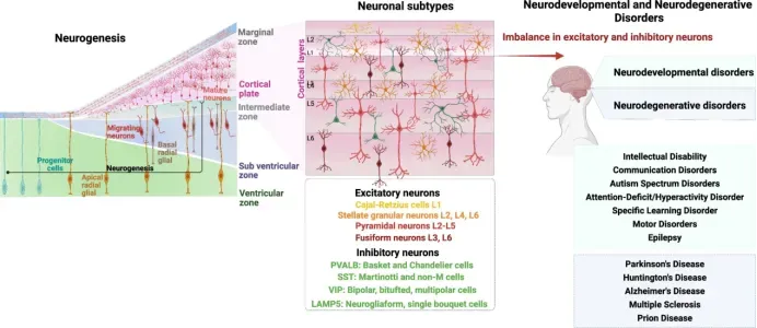
Neurogenesis during fetal brain development, cortical neuronal subtypes, and associated neurodevelopmental and neurodegenerative disorders (PMID: 39095595).
Disruption of brain developmental programs or the balance between neuronal populations can contribute to neurodevelopmental and neurodegenerative disorders. We provide validated antibody solutions for comprehensive profiling of neurodegenerative disease mechanisms—from pathological protein aggregation and proteostasis failure to neuronal degeneration, synaptic dysfunction, neuroinflammation, and cellular stress.
Integrated molecular and spatial profiling enables researchers to connect pathological hallmarks with cellular states and functional consequences across disease initiation, progression, and therapeutic response.
Neurodegenerative Disease Research Portfolio
| Disease Model | Major Molecular Pathology | Representative & Emerging Targets |
|---|---|---|
| Alzheimer’s Disease (AD) | Aβ plaques, Tau pathology & neuroimmune dysfunction | APP, Aβ, BACE1, PSEN1, Tau, p-Tau (Ser202/Thr205), p-Tau (Thr181), TREM2, APOE, CD33, GPNMB, LPL, C1q, C3, GFAP, IBA1, PSD95, Synaptophysin, NeuN |
| Parkinson’s Disease (PD) | α-Synuclein pathology & dopaminergic neuron degeneration | α-Synuclein, p-α-Syn (Ser129), TH, DAT, VMAT2, LRRK2, GBA1/GCase, PINK1, Parkin, DJ-1, LAMP2A, TFEB, TOM20, NLRP3, NeuN |
| ALS / Frontotemporal Dementia (ALS/FTD) | TDP-43/FUS proteinopathy & motor/cortical neuron degeneration | TDP-43, p-TDP-43, FUS, C9orf72, STMN2, UNC13A, TBK1, OPTN, SQSTM1/p62, NEFH, ChAT, ATXN2, Nucleoporins |
| Multiple Sclerosis (MS) | Demyelination, chronic CNS inflammation & axonal degeneration | MBP, MOG, PLP1, OLIG2, SOX10, PDGFRA, NG2/CSPG4, IBA1, TMEM119, GFAP, C3, C1q, CD3, CD4, CD8, BTK, LINGO-1, GPX4 |
| Brain Aging & Neurodegeneration | Cellular senescence, proteostasis decline & chronic neuroinflammation | p16INK4a, p21, γH2AX, Lamin B1, GLB1, SASP factors, cGAS, STING, NLRP3, NRF2, SIRT1, AMPK, mTOR, LC3B, p62/SQSTM1, TFEB, GPX4, ACSL4, 4-HNE |
Featured Products
| Catalog No. | Product Name | Reactivity | Validated Applications |
|---|---|---|---|
| WR5246 | U-Blot® Amyloid-β (1-42) Rabbit mAb | Human, Mouse | WB, IHC, IF, ELISA |
| WR4293 | U-Blot® Amyloid-β Rabbit mAb | Human, Mouse, Rat, Hamster | WB, IHC, IF, IP, ELISA |
| WR4620 | U-Blot® ApoE Rabbit mAb | Human, Mouse, Rat | WB, IHC, IF, IP, ELISA |
| WR4095 | U-Blot® Cathepsin D Rabbit mAb | Human, Mouse, Rat | WB, IHC, IF, IP, ELISA |
| WR4667 | U-Blot® eIF2α (Phospho Ser51) Rabbit mAb | Human, Mouse, Rat, Chicken, Dog, Pig, Fish | WB, IHC, IF, ELISA |
| WR4287 | U-Blot® eIF2α Rabbit mAb | Human, Mouse, Rat, Fish | WB, IHC, IF, IP, ELISA |
| WR5278 | U-Blot® GBA Rabbit mAb | Human, Rat | WB, IHC, IF, ELISA |
| WR4151 | U-Blot® GFAP Rabbit mAb | Human, Mouse, Rat | WB, IHC, IF, IP, ELISA |
| WR8058 | U-Blot® Glial Fibrillary Acidic Protein (GFAP) Rabbit mAb (Ready to Use) | Human, Rat | IHC |
| WR4181 | U-Blot® GRP78/BiP Rabbit mAb | Human, Mouse, Rat, Fish | WB, IHC, IF, ELISA |
| WR4118 | U-Blot® Hsp70 Rabbit mAb | Human, Mouse, Rat | WB, IHC, IF, ELISA |
| WR5736 | U-Blot® Huntingtin Rabbit mAb | Human, Mouse, Rat | WB, IHC, IF, ELISA |
| WR4144 | U-Blot® Iba1 Rabbit mAb | Human, Mouse, Rat | WB, IHC, IF, IP, ELISA |
| WR4040 | U-Blot® LAMP1 Rabbit mAb | Human | WB, IHC, IF, ELISA |
| WR4136 | U-Blot® LAMP2 Rabbit mAb | Human, Mouse, Rat | WB, IHC, IF, ELISA |
| WR4126 | U-Blot® LC3B Rabbit mAb | Human, Mouse, Rat, Pig | WB, IHC, IF, ELISA |
| WR4820 | U-Blot® LRRK2 Rabbit mAb | Human, Mouse, Rat | WB, IHC, IF, IP, ELISA |
| WR4578 | U-Blot® MAP2 Rabbit mAb | Human, Mouse, Rat | WB, IHC, IF, ELISA |
| WR4916 | U-Blot® Neurofilament (NF-L) Rabbit mAb | Human, Mouse, Rat | WB, IHC, IF, IP, ELISA |
| WR4138 | U-Blot® Neurofilament heavy polypeptide Rabbit mAb | Human, Mouse, Rat | WB, IHC, IF, ELISA |
| WR8087 | U-Blot® Neurofilament(NF-L) Rabbit mAb (Ready to Use) | Human | IHC |
| WR5210 | U-Blot® NF-M (Phospho Ser614/Ser619) Rabbit mAb | Human, Mouse, Rat | WB, IHC, IF, ELISA |
| WR8094 | U-Blot® PGP 9.5 Rabbit mAb (Ready to Use) | Human, Mouse, Rat | IHC |
| WR4197 | U-Blot® PGP9.5 Rabbit mAb | Human, Mouse, Rat | WB, IHC, IF, IP, ELISA |
| WR5906 | U-Blot® SIRT2 Rabbit mAb | Human, Mouse, Rat | WB, IHC, IF, IP, ELISA |
| WR4045 | U-Blot® SOD1 Rabbit mAb | Human, Mouse, Rat | WB, IHC, IF, ELISA |
| WR4790 | U-Blot® SQSTM1/p62 (Phospho Ser349) Rabbit mAb | Human, Mouse, Rat | WB, IHC, IF, ELISA |
| WR4007 | U-Blot® SQSTM1/p62 Rabbit mAb | Human, Mouse, Rat | WB, IHC, IF, IP, ELISA |
| WR4840 | U-Blot® Synuclein-α (Phospho Ser129) Rabbit mAb | Human, Mouse, Rat | WB, IF, IP, ELISA |
| WR4587 | U-Blot® Synuclein-α Rabbit mAb | Human, Mouse, Rat, C.elegans | WB, IHC, IF, IP, ELISA |
| WR4621 | U-Blot® Tau (Phospho Ser396) Rabbit mAb | Human, Mouse, Rat | WB, IHC, IF, ELISA |
| WR4576 | U-Blot® Tau Rabbit mAb | Human, Mouse, Rat | WB, IHC, IF, IP, ELISA |
| WR4049 | U-Blot® TDP43 Rabbit mAb | Human, Mouse, Rat | WB, IHC, IF, IP, ELISA |
| WR4160 | U-Blot® TLS/FUS Rabbit mAb | Human, Mouse, Rat | WB, IHC, IF, ELISA |
| WR4934 | U-Blot® TREM2 Rabbit mAb | Human | WB, IF, IP, ELISA, IHC |
| WR4274 | U-Blot® UBA52 Rabbit mAb | Human, Mouse, Rat | WB, IHC, IF, IP, ELISA |
| WR4240 | U-Blot® Ubiquitin K48 Rabbit mAb | Human, Mouse, Rat | WB, IHC, IF, ELISA |
| WR4944 | U-Blot® Ubiquitin K63 Rabbit mAb | Human, Mouse, Rat | WB, IHC, IF, ELISA |
| WR4167 | U-Blot® XBP1 Rabbit mAb | Human, Mouse, Rat | WB, IHC, IF, IP, ELISA |
| WR4338 | U-Blot® β3 Tubulin Rabbit mAb | Human, Mouse, Rat | WB, IHC, IF, IP, ELISA |
Alzheimer’s Disease Molecular Research Workflow
From Pathological Hallmarks to Cellular Mechanisms
Alzheimer’s disease involves interconnected alterations in protein homeostasis, mitochondrial function, cellular aging, neuroimmune signaling, synaptic integrity, and neuronal survival. Integrated antibody profiling enables researchers to connect pathological hallmarks with molecular mechanisms and cellular consequences across multiple biological scales.
01 | Proteostasis Dysfunction
Decipher Protein Quality Control and Clearance Failure
Key Biological Processes
Protein Misfolding→Aggregation→Autophagy–Lysosome Dysfunction→Impaired Clearance→Proteotoxic Stress
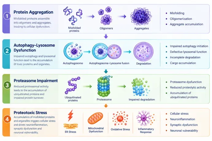
Neuronal function depends on tightly regulated protein homeostasis. Disruption of protein folding, trafficking, autophagy–lysosome pathways, or ubiquitin–proteasome-mediated degradation can promote pathological protein accumulation and proteotoxic stress, contributing to synaptic dysfunction and neuronal vulnerability.
Integrated profiling of proteinopathy and protein quality-control pathways enables investigation of how impaired proteostasis contributes to neurodegenerative disease progression.
Representative Targets
| Functional Module | Key Targets |
|---|---|
| Proteinopathy | Aβ, Tau, p-Tau |
| Autophagy | LC3B, SQSTM1/p62, ATG5, ATG7 |
| Lysosomal Function | LAMP1, LAMP2, CTSD |
| Autophagy Regulation | TFEB, ULK1, Beclin-1 |
| Ubiquitin–Proteasome System | Ubiquitin, Proteasome subunits, p62 |
02 | Mitochondrial Stress
Defining Mitochondrial Quality Control, Bioenergetics & Oxidative Stress
Key Biological Processes
Mitochondrial Dysfunction→OXPHOS Impairment→ROS Accumulation→Mitophagy / Quality-Control Response→Energy Failure→Neuronal Vulnerability
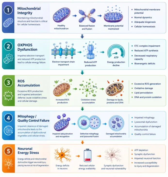
Neurons have exceptionally high energetic demands and depend on robust mitochondrial function to maintain membrane potential, synaptic activity, and cellular survival. Mitochondrial dysfunction can impair oxidative phosphorylation, increase oxidative stress, and disrupt mitochondrial quality-control pathways, including mitophagy and biogenesis.
Profiling mitochondrial integrity, bioenergetic pathways, oxidative stress, and quality-control mechanisms provides insight into their contribution to synaptic dysfunction and neurodegeneration.
Representative Targets
| Functional Module | Key Targets |
|---|---|
| Mitochondrial Structure | TOM20, VDAC1, HSP60 |
| OXPHOS | COX IV, ATP5A, SDHB, NDUFB8 |
| Mitochondrial Stress | HSP60, 4-HNE, ROS-associated markers |
| Mitophagy | PINK1, Parkin, BNIP3, BNIP3L/NIX |
| Mitochondrial Biogenesis | PGC-1α, NRF1, TFAM |
| Antioxidant Response | NRF2, SOD2, GPX4 |
03 | Cellular Aging
Decoding Senescence, DNA Damage & Inflammatory Remodeling
Research Focus
DNA Damage→Persistent DDR→Cellular Senescence→SASP→Chronic Inflammatory Signaling→Neural Dysfunction
Key Biological Processes
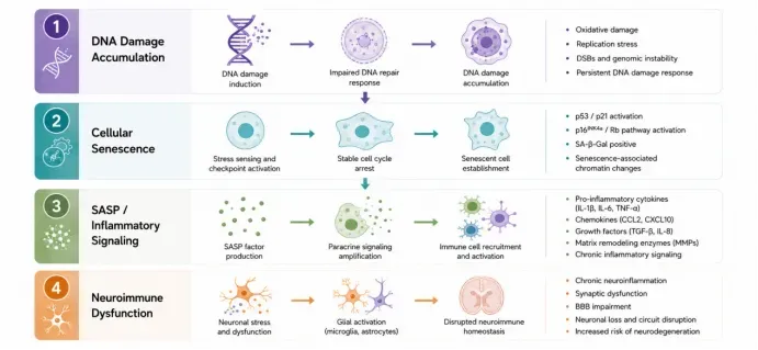
Accumulation of DNA damage and persistent stress responses can promote cellular senescence and senescence-associated secretory phenotypes (SASP), reshaping the neural microenvironment through chronic inflammatory signaling. These changes may contribute to glial dysfunction, synaptic impairment, vascular alterations, and neuronal vulnerability during aging and neurodegenerative disease.
Representative Targets
| Functional Module | Key Targets |
|---|---|
| Cell-Cycle Arrest | p16INK4a, p21 |
| DNA Damage Response | γH2AX, 53BP1 |
| Nuclear Integrity | Lamin B1 |
| Senescence-associated Activity | GLB1 |
| SASP / Inflammation | IL-6, IL-1β, NF-κB |
| Innate Immune Activation | cGAS, STING |
| Stress Response | NRF2, SIRT1 |
Integrated Alzheimer’s Disease Research Panel
Multi-Dimensional Profiling of Pathology, Cell States & Neural Dysfunction
| Research Question | Recommended Marker Panel |
|---|---|
| Amyloid pathology | APP + Aβ + BACE1 + PSEN1 |
| Tau pathology | Total Tau + p-Tau (Ser202/Thr205) + p-Tau (Thr181) + MAP2 |
| Microglial activation / state transition | IBA1 + TMEM119 + P2RY12 + CD68 |
| Disease-associated microglia | TREM2 + APOE + GPNMB |
| Astrocyte reactivity | GFAP + ALDH1L1 + C3 + Vimentin |
| Neuroinflammatory signaling | NLRP3 + C1q + C3 + NF-κB |
| Synaptic dysfunction | PSD95 + Synaptophysin + SNAP25 + Synapsin I |
| Neuronal integrity | NeuN + MAP2 + NF-L/NF-H |
| Proteostasis dysfunction | LC3B + p62 + LAMP1 + CTSD + TFEB |
| Cellular senescence / aging | p16 + p21 + γH2AX + Lamin B1 |
| Mitochondrial stress | TOM20 + COX IV + ATP5A + PINK1/Parkin |
| Ferroptosis / oxidative stress | GPX4 + ACSL4 + 4-HNE + NRF2 |
Neuronal Proteostasis & Protein Clearance(Human Glioma FFPE)
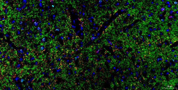

This panel enables simultaneous spatial profiling of protein aggregation and autophagy–lysosomal pathways in human glioma tissue, supporting research on neurodegenerative disease mechanisms and pathological protein clearance.