
info@mail.ucallm.com
Neuroimmune Landscape
Decoding Immune Regulation and Glial State Dynamics in the Brain
From Glial States to Cellular Interactions, Spatial Context & Disease Mechanisms
The CNS is an actively regulated immune environment in which neurons, microglia, astrocytes, and other glial populations interact dynamically to maintain homeostasis and respond to pathological stress.
Integrated neuroimmune profiling enables researchers to characterize glial states, inflammatory signaling, cellular interactions, and neuronal consequences across health, aging, injury, and neurodegenerative disease.
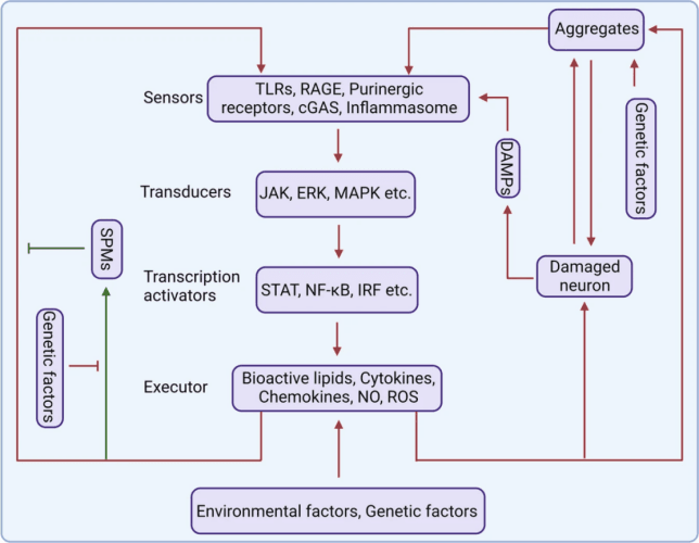
The role of inflammation in neurodegenerative (PMID: 37433768).
Key Molecular Targets
| Pathological Stage | Key Molecular Targets / Markers |
|---|---|
| Pathological Stimulus | Aβ, p-Tau, α-Synuclein |
| Microglial Activation | IBA1/AIF1, TMEM119, P2RY12, CD68 |
| Inflammatory Signaling | NF-κB, NLRP3, p38 MAPK, JNK, ERK1/2 |
| Inflammatory Mediators | IL-1β, TNF-α, IL-6, CCL2 |
| Astrocyte Reactivity | GFAP, S100B, Vimentin |
| Neuroimmune Crosstalk | C3, C1q, TGF-β, CX3CL1/CX3CR1 |
| Synaptic Dysfunction | PSD95, Synaptophysin, SNAP25, Homer1 |
| Neuronal Stress / Injury | NeuN, MAP2, NF-L, APP |
| Neuronal Degeneration | Cleaved Caspase-3, BAX, BCL-2 |
Featured Products
| Catalog No. | Product Name | Reactivity | Validated Applications |
|---|---|---|---|
| WR5176 | U-Blot® ALDH1L1 Rabbit mAb | Human, Mouse, Rat | WB, IHC, IF, IP, ELISA |
| WR4902 | U-Blot® AQP1 Rabbit mAb | Human, Mouse, Rat | WB, IHC, IF, ELISA |
| WR5094 | U-Blot® AQP4 Rabbit mAb | Human, Mouse, Rat | WB, IHC, IF, IP, ELISA |
| WR4225 | U-Blot® CD146 Rabbit mAb | Human, Mouse, Rat | WB, IHC, IF, ELISA |
| WR4322 | U-Blot® CD206/MRC1 Rabbit mAb | Human, Mouse, Rat | WB, IHC, IF, IP, ELISA |
| WR4185 | U-Blot® CD31 Rabbit mAb | Human, Mouse, Rat | WB, IHC, IF, ELISA |
| WR4340 | U-Blot® CD68 Rabbit mAb | Mouse, Rat | WB, IHC, IF, ELISA |
| WR8034 | U-Blot® CD68 Rabbit mAb (Ready to Use) | Human | IHC |
| WR5184 | U-Blot® Claudin-11 Rabbit mAb | Human, Mouse, Rat | WB, IF, ELISA |
| WR5307 | U-Blot® EAAT2 Rabbit mAb | Human, Mouse, Rat | WB, IHC, IF, ELISA |
| WR4151 | U-Blot® GFAP Rabbit mAb | Human, Mouse, Rat | WB, IHC, IF, IP, ELISA |
| WR8058 | U-Blot® Glial Fibrillary Acidic Protein (GFAP) Rabbit mAb (Ready to Use) | Human, Rat | IHC |
| WR4010 | U-Blot® Glutamine Synthetase Rabbit mAb | Human, Mouse, Rat | WB, IHC, IF, ELISA |
| WR4144 | U-Blot® Iba1 Rabbit mAb | Human, Mouse, Rat | WB, IHC, IF, IP, ELISA |
| Catalog No. | Product Name | Reactivity | Validated Applications |
|---|---|---|---|
| WR4262 | U-Blot® LYVE1 Rabbit mAb | Human, Mouse, Rat | WB, IHC, IF, ELISA |
| WR8081 | U-Blot® Myelin Basic Protein (MBP) Rabbit mAb (Ready to Use) | Human, Mouse, Rat | IHC |
| WR4290 | U-Blot® OCLN Rabbit mAb | Human, Mouse, Rat | WB, IHC, IF, IP, ELISA |
| WR4617 | U-Blot® OLIG2 Rabbit mAb | Human, Mouse, Rat | WB, IHC, IF, ELISA |
| WR8089 | U-Blot® OLIG2 Rabbit mAb (Ready to Use) | Human, Mouse | IHC |
| WR5120 | U-Blot® P2Y12 Rabbit mAb | Human, Mouse, Rat | WB, IF, IP, ELISA, IHC |
| WR4375 | U-Blot® PDGFR-α Rabbit mAb | Human, Mouse, Rat | WB, IF, IP, ELISA |
| WR4385 | U-Blot® PDGFR-β Rabbit mAb | Human, Mouse, Rat | WB, IHC, IF, IP, ELISA |
| WR5249 | U-Blot® PLP Rabbit mAb | Human, Mouse, Rat | WB, IHC, IF, ELISA |
| WR4271 | U-Blot® S100B Rabbit mAb | Human, Mouse, Rat | WB, IHC, IF, ELISA |
| WR8113 | U-Blot® S100B Rabbit mAb (Ready to Use) | Human, Mouse, Rat | IHC |
| WR4425 | U-Blot® SOX10 Rabbit mAb | Human, Mouse, Rat | WB, IHC, IF, ELISA, IP |
| WR4934 | U-Blot® TREM2 Rabbit mAb | Human | WB, IF, IP, ELISA, IHC |
| WR4346 | U-Blot® VE Cadherin Rabbit mAb | Mouse, Rat | WB, IHC, IF, IP, ELISA |
| WR5139 | U-Blot® VE-Cadherin Rabbit mAb | Human | WB, IF, IP, ELISA |
| WR4166 | U-Blot® VWF Rabbit mAb | Human, Mouse, Rat | WB, IHC, IF, IP, ELISA |
| WR4021 | U-Blot® α smooth muscle actin Rabbit mAb | Human, Mouse, Rat | WB, IHC, IF, IP, ELISA |
Microglia State Profiling
Resolving Microglial Identity, Functional State & Disease-Associated Remodeling
Microglia are the principal immune-surveillance cells of the CNS. Their phenotypic states are dynamically shaped by developmental cues, tissue environment, injury, aging, and disease-associated signals.
Multiparameter profiling enables researchers to distinguish homeostatic, reactive, phagocytic, and disease-associated phenotypes while linking microglial remodeling to neuronal and synaptic dysfunction.
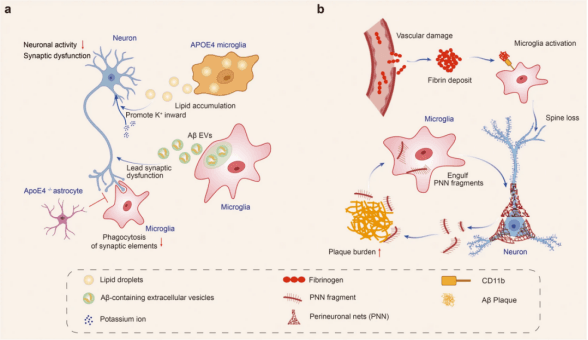
Dysfunctional microglia impair neuronal activity in Alzheimer's disease (PMID:37735487).
Microglia Marker Panel
| Microglial State | Biological Function | Representative Markers |
|---|---|---|
| Microglial Identity | Microglia identification and localization | IBA1/AIF1, TMEM119 |
| Homeostatic Microglia | Immune surveillance, tissue maintenance, synaptic support | TMEM119, P2RY12, CX3CR1 |
| Reactive Microglia | Injury sensing, inflammatory activation, environmental response | IBA1/AIF1, CD68, MHC-II |
| Phagocytic Microglia | Phagocytosis, debris clearance, cellular remodeling | CD68, TREM2, LAMP1 |
| Disease-Associated Microglia | Neurodegenerative response, lipid remodeling, disease adaptation | TREM2, APOE, GPNMB, LPL |
| Inflammatory Microglia | Cytokine production and inflammatory amplification | IL-1β, TNF-α, NLRP3, NF-κB |
| Neurodegeneration-Associated State | Synaptic remodeling, neuronal stress and disease progression | TREM2, APOE, GPNMB, CD68 |
Astrocyte State Profiling
From Homeostatic Support to Reactive Remodeling and Neuroinflammatory Responses
Astrocytes are multifunctional regulators of CNS homeostasis, contributing to neuronal metabolic support, neurotransmitter and ion homeostasis, synaptic regulation, neurovascular function, and responses to injury and inflammation.
Profiling astrocyte identity and state provides molecular insight into how astrocyte remodeling influences neural function and disease progression.
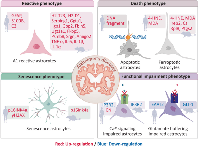
The pathological phenotypes of astrocytes in human and mouse AD (PMID: 38172603).
Astrocyte Marker Panel
| Functional Category | Representative Markers | Key Biological Function |
|---|---|---|
| Astrocyte Identity | ALDH1L1, GFAP, SOX9, S100B | Astrocyte identification |
| Homeostatic State | ALDH1L1, AQP4, S100B | Tissue and metabolic homeostasis |
| Reactive State | GFAP, Vimentin, STAT3 | Reactive remodeling and injury response |
| Inflammatory State | C3, NF-κB, STAT3, SERPING1 | Neuroinflammatory signaling |
| Metabolic Support | GLT-1/SLC1A2, GLAST/SLC1A3, GS/GLUL | Glutamate clearance and metabolic coupling |
| Water / Ion Homeostasis | AQP4, KCNJ10/Kir4.1 | Water, potassium and extracellular homeostasis |
| Cell-Cell Communication | Connexin43/GJA1, THBS1, SPARCL1 | Gap-junction and synaptic regulation |
| Neurodegeneration-Associated State | C3, APOE, SERPINA3, GFAP | Disease-associated remodeling and neuronal dysfunction |
Neuroimmune Research Applications
Profiling Cellular Interactions Across Brain Health and Disease
| Research Area | Key Research Questions |
|---|---|
| Neuroinflammation | How are inflammatory pathways activated and sustained? |
| Neurodegeneration | How do disease-associated glial states contribute to neuronal vulnerability? |
| Brain Aging | How does aging reshape glial homeostasis and inflammatory responses? |
| Neural Injury & Repair | How do glial responses influence tissue remodeling and recovery? |
Representative Applications
Microglial State Profiling · Astrocyte Reactivity · Neuroimmune Signaling · Proteinopathy-Driven Inflammation · Synaptic Remodeling · Neuronal Vulnerability
Integrated Neuroimmune Profiling Workflow
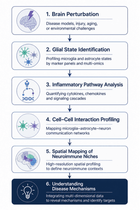
Our integrated neuroimmune research solutions connect cellular profiling, signaling analysis, cell–cell interaction, and spatial mapping to provide a multidimensional view of neuroimmune regulation and disease mechanisms.
Validated Results
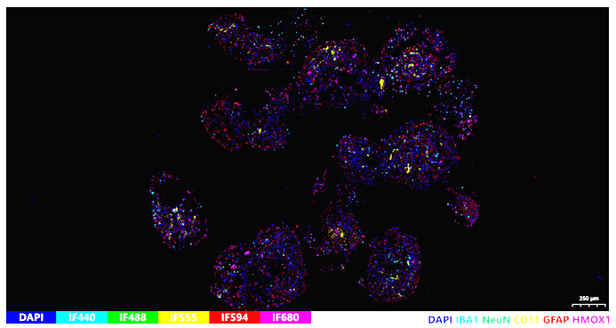
 |  |  |
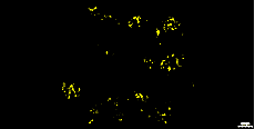 | 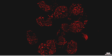 | 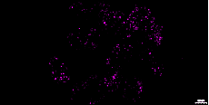 |
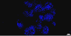
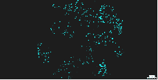
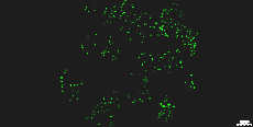
Neural organoid models combined with advanced fluorescence imaging enable visualization of cellular composition, differentiation, morphology and functional responses in 3D.
Products for Organoid Imaging
| Target | Product Name | Catalog No. | Reactivity | Applications | Biological Interpretation |
|---|---|---|---|---|---|
| NeuN (RBFOX3) | U-Blot® NeuN Rabbit mAb | WR4580 | Human, Mouse, Rat | WB, IHC, IF, ELISA | Identifies mature neurons and evaluates neuronal survival and neurodegeneration after hemorrhagic injury. |
| GFAP | U-Blot® GFAP Rabbit mAb | WR4151 | Human, Mouse, Rat | WB, IHC, IF, ELISA | Detects reactive astrocytes and measures astrogliosis surrounding the hematoma and injured tissue. |
| IBA1 | U-Blot® Iba1 Rabbit mAb | WR4144 | Human, Mouse, Rat | WB, IHC, IF, ELISA | Assesses microglial activation, neuroinflammatory responses, and immune-cell accumulation after ICH. |
| CD31 | U-Blot® CD31 Rabbit mAb | WR4185 | Human, Mouse, Rat | WB, IHC, IF, ELISA | Labels cerebral blood vessels to evaluate vascular integrity, angiogenesis, and blood–brain barrier alterations. |
| HO-1/HMox1 | U-Blot® HO-1 Rabbit mAb | WR4310 | Human, Mouse, Rat | WB, IHC, IF, ELISA | Measures oxidative stress and heme degradation induced by hemorrhage, reflecting antioxidant and injury responses. |