
info@ucallmlabs.com
1. Introduction
Cardiovascular diseases have long remained the leading cause of mortality worldwide. Traditional cardiac research models have significant limitations: animal models differ greatly from humans in cardiac development and genetic regulatory pathways, resulting in low accuracy in predicting drug‑induced cardiotoxicity; human primary cardiomyocytes are difficult to obtain and cannot be expanded on a large scale, making them unsuitable for supporting high‑throughput drug screening, genetic disease modeling, and regenerative medicine development. Human pluripotent stem cells (hPSCs, including human embryonic stem cells hESCs and patient‑derived induced pluripotent stem cells hiPSCs) possess unlimited proliferation capacity and the unique ability to differentiate into all types of human cardiovascular cells, making them a core cellular resource for constructing "in vitro mini‑heart" models and advancing innovative cardiovascular research and development.
Human embryonic heart development is precisely regulated by the sequential coordination of signaling pathways including Activin/Nodal, BMP, Wnt, and VEGF. The precise concentration and regulatory timing of these signals directly determine whether mesodermal cells differentiate toward cardiovascular or hematopoietic lineages. Classic authoritative studies have demonstrated that different human stem cell lines exhibit significant differences in endogenous Nodal and BMP basal expression levels, making it impossible to achieve efficient cardiac differentiation using a universal cytokine formulation. HES2 and H1 human embryonic stem cells exhibit relatively weak endogenous signaling activity and require exogenous supplementation with Activin A (3–6 ng/mL) combined with BMP4 (10 ng/mL) to induce cardiac mesoderm formation; whereas the MSC‑iPS1 induced pluripotent stem cell line exhibits high endogenous BMP4 expression, and additional Activin A supplementation completely inhibits cardiovascular progenitor generation, requiring only BMP4 (2–10 ng/mL) for successful induction. All cell lines require continuous supplementation with bFGF and VEGF to maintain progenitor cell survival.
Based on this stage‑specific signal‑optimized differentiation technology, we can stably and efficiently produce human cardiovascular lineage cells, including human cardiomyocytes and vascular smooth muscle cells, to serve three major application areas: first, preclinical cardiac toxicity screening for new drug development, significantly reducing dependence on animal experiments and improving the accuracy of human response prediction; second, construction of in vitro disease models for genetic cardiomyopathies and arrhythmias, enabling personalized investigation of pathological mechanisms; and third, cardiac regenerative medicine and tissue engineering‑based preparation of cardiac seed cells, providing highly biomimetic and standardized human cell platforms for cardiovascular innovative drug development and cell therapy research. This approach aligns with the global trend among pharmaceutical companies and regulatory agencies toward reducing animal experimentation and promoting the application of human‑based in vitro models.
2. Workflow Overview
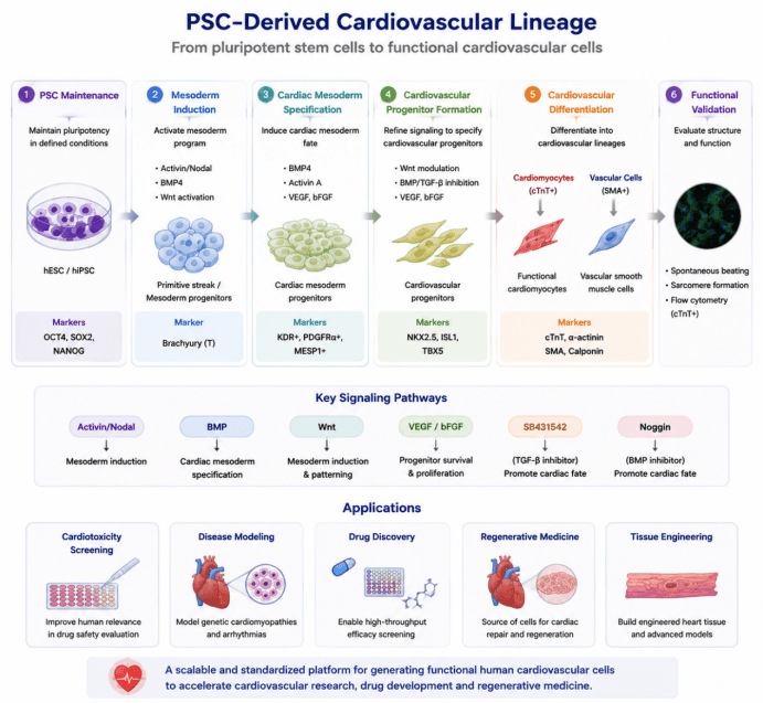
By regulating signaling pathways including Activin/Nodal, BMP, Wnt, and VEGF in a stage‑specific manner, hESCs/hiPSCs undergo mesoderm induction, cardiac mesoderm specification, and cardiovascular progenitor formation, ultimately generating functional cardiomyocytes and vascular cells. PSC‑derived cardiovascular cells provide a standardized human cell platform for cardiac toxicity evaluation, disease model construction, drug screening, and regenerative medicine research.
3. Differentiation Protocol
| Differentiation Stage | Added Factors / Culture Conditions | Core Biological Function | Corresponding Cell Type / Developmental Stage | Stage‑Specific Identification Markers |
|---|---|---|---|---|
| Pluripotent Stem Cell Maintenance and Preparation (hESC/hiPSC) | Matrigel coating; E8/mTeSR complete medium; Y‑27632 supplementation during cell passaging | Maintains pluripotency, reduces single‑cell apoptosis, improves cell attachment efficiency, and eliminates feeder cell contamination | Human pluripotent stem cells (hPSCs) | OCT4⁺, SOX2⁺, NANOG⁺ |
| Stage 1: Embryoid Body (EB) Initiation Culture (Differentiation D0–D4) | Serum‑free differentiation medium without exogenous growth factors; suspension culture to form embryoid bodies (EBs) | Initiates primitive streak and mesoderm differentiation programs, activates endogenous Wnt signaling, and confers mesodermal differentiation potential | Early embryoid bodies; primitive streak precursor cells | Upregulation of Brachyury (T) expression |
| Stage 2: Cardiac Mesoderm Induction (Differentiation D4–D5 Detection) | Basic factors: bFGF 5 ng/mL, VEGF 5 ng/mL. Optimal for HES2/H1 cell lines: Activin A 3–6 ng/mL + BMP4 10 ng/mL. Optimal for MSC‑iPS1 cell line: BMP4 2–10 ng/mL without Activin A supplementation | Precisely regulates Activin/Nodal and BMP signaling gradients to induce cardiac‑specific mesoderm. High concentrations of BMP4 inhibit cardiovascular lineage specification and promote hematopoietic mesoderm fate | Cardiac mesoderm progenitors (cardiovascular progenitor cells) | KDR⁺PDGFR‑α⁺ (K⁺P⁺); strong expression of transcription factor MESP1 |
| Stage 3: Cardiac Lineage Specification (Critical Regulation Window at Differentiation D3) | SB‑431542 (blocking Activin/Nodal signaling) ± Dorsomorphin/Noggin (blocking BMP signaling); maintenance with VEGF, bFGF, and DKK1 | Suppresses continuous endogenous Nodal/BMP signaling, removes inhibition of cardiomyocyte differentiation, and stabilizes cardiac cell fate commitment (essential step for human iPSC differentiation) | Committed cardiovascular progenitors with loss of hematopoietic differentiation potential | Sustained upregulation of NKX2.5, ISL1, TBX5, MEF2C; significant downregulation of hematopoietic marker SCL |
| Stage 4: Monolayer Cardiomyocyte Expansion and Maturation (Differentiation D5–D20) | StemPro‑34 serum‑free cardiomyocyte medium; supplemented with L‑glutamine, ascorbic acid, VEGF, bFGF, FGF10, and DKK1; gelatin‑coated plates for adherent monolayer culture | Promotes cardiovascular progenitor proliferation and terminal differentiation into cardiomyocytes and vascular smooth muscle cells; spontaneous beating and formation of cardiac cell sheets | Terminal cardiovascular lineage: cardiomyocytes and vascular smooth muscle cells | Cardiomyocyte markers: cTnT⁺. Vascular smooth muscle markers: SMA⁺, Calponin. Mature cardiomyocyte markers: MLC2A, MLC2V, NPPA. Low‑level DDR2 expression (fibroblast marker) |
| Hematopoietic Control Lineage (Non‑Cardiovascular) | Low concentration Activin A + high concentration BMP4 induction | Induces hematopoietic mesoderm differentiation as a negative control for cardiovascular lineage specification | Hematopoietic mesoderm and hematopoietic progenitor cells | KDR⁺PDGFR‑α⁻ (K⁺P⁻); CD41⁺, high SCL expression; absence of MESP1 expression |
| Functional Validation Stage | Continuous cardiomyocyte monolayer culture; intracellular flow cytometry, immunofluorescence staining, and microscopic observation of beating activity | Validates cardiomyocyte contractile function, sarcomeric structure, and quantitatively evaluates cardiomyocyte purity | Functional beating cardiomyocytes and composite cardiovascular cell sheets | Sarcomeric α‑actinin; flow cytometric quantification of cTnT‑positive cell percentage |