
info@ucallmlabs.com
The clinical demand for end-stage liver failure, hereditary metabolic liver diseases, viral hepatitis, and drug-induced liver injury continues to increase, while donor liver resources remain extremely limited, restricting the application of liver transplantation and hepatocyte transplantation. Primary human hepatocytes are essential resources for hepatotoxicity screening, liver development studies, in vitro liver disease modeling, and regenerative medicine research. However, primary hepatocytes rely on donor liver availability, rapidly dedifferentiate and lose drug-metabolizing functions during in vitro culture, and cannot be stably expanded on a large scale, greatly limiting drug discovery and regenerative medicine development.
In 2010, a landmark serum-free differentiation protocol was reported in Hepatology, establishing a standardized system that faithfully recapitulates human liver embryonic development. This four-step, two-dimensional monolayer-directed differentiation system was developed for human embryonic stem cells (hESCs, H9) and induced pluripotent stem cells (hiPSCs). The entire process avoids embryoid body formation, animal serum, and feeder cells. Through sequential regulation of five key signaling molecules, including Activin A, BMP4, FGF2, HGF, and OSM, combined with precise hypoxic culture conditions, high-purity hepatocyte-like cells can be generated within approximately 20 days, with albumin-positive cells accounting for more than 80%. Multiple functional assays demonstrated that the induced cells exhibit typical hepatocyte physiological functions, including glycogen storage, lipid metabolism, low-density lipoprotein uptake, indocyanine green clearance, and urea synthesis. These cells also highly express liver-specific markers such as albumin, HNF4α, and cytochrome P450 drug-metabolizing enzymes. In vivo transplantation studies further confirmed that these hepatocyte-like cells can stably engraft into mouse liver parenchyma, continuously secrete human albumin, and possess liver regenerative potential. This differentiation platform can generate multiple liver cell types, including hepatocytes, liver sinusoidal endothelial cells, and Kupffer cells. This fully defined, highly reproducible, and scalable differentiation system provides a standardized human hepatic cell platform. It can be widely applied in high-throughput hepatotoxicity screening of innovative drugs, patient-specific liver disease modeling, studies of human liver embryonic development, hepatocyte transplantation cell source development, and artificial liver biomaterial research, providing stable, compliant, and functionally mature human hepatic cell resources for liver-related basic research and preclinical regenerative medicine development.
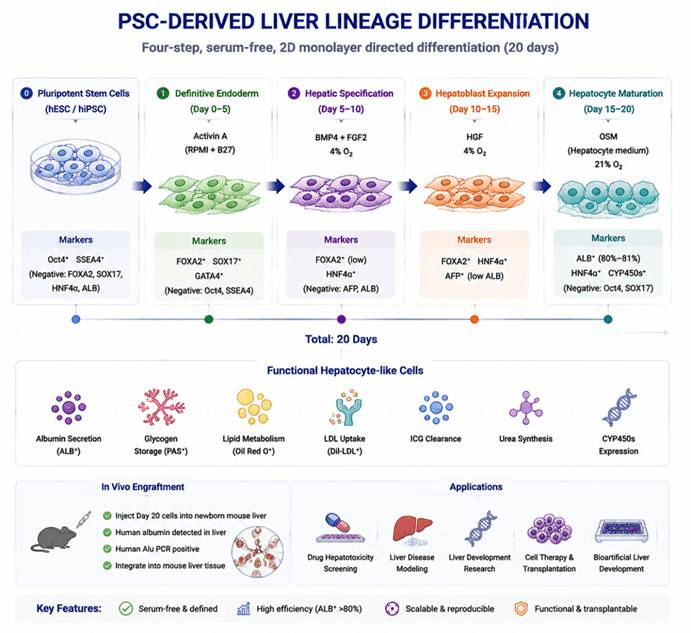
The differentiation process of pluripotent stem cells toward functional hepatocyte-like cells. hESCs/hiPSCs are first induced by Activin A to form definitive endoderm, followed by hepatic lineage specification under BMP4/FGF2 stimulation. Hepatoblast expansion is promoted by HGF, and the cells are finally matured into functional hepatocyte-like cells through OSM induction. The derived cells express hepatocyte markers including ALB, HNF4α, and CYP450, and exhibit hepatic functions such as albumin secretion, metabolism, and drug biotransformation, providing a valuable platform for liver disease modeling, drug screening, and regenerative medicine research.
Differentiation Stage | Added Factors / Culture Conditions | Core Biological Function | Corresponding Cell Type / Developmental Stage | Stage-Specific Identification Markers |
Pluripotent Stem Cell Maintenance (Starting Cells) | Matrigel-coated plates; MEF-conditioned stem cell medium; 4% hypoxic environment; no induction cytokines; human H9 ES cells / human dermal fibroblast-derived hiPSCs | Maintains Oct4 and SSEA4 pluripotent state and provides homogeneous starting cells | Undifferentiated human pluripotent stem cells (ES/hiPSCs) | Positive: Oct4, SSEA4Negative: FOXA2, SOX17, HNF4α, AFP, ALB |
Stage 1: Definitive Endoderm Induction (Day 0–5) | Basal medium: RPMI + B27; 100 ng/mL Activin A; normoxic condition (5% CO₂); 2D monolayer culture | Mimics embryonic gastrulation and efficiently induces definitive endoderm while avoiding yolk sac visceral endoderm contamination | Embryonic definitive endoderm (upstream progenitor of liver development) | Positive: FOXA2, SOX17, GATA4Negative: Oct4, SSEA4, HNF4α (visceral endoderm marker) |
Stage 2: Hepatic Specification (Day 5–10) | RPMI + B27; 20 ng/mL BMP4 + 10 ng/mL FGF2; continuous 4% hypoxic environment | Recapitulates septum transversum mesenchyme signaling during embryonic development, directing endoderm toward hepatic lineage and suppressing intestinal endoderm differentiation | Early hepatic specified progenitor cells (hepatic fate-committed cells) | Maintained: FOXA2Downregulated: GATA4Newly expressed: HNF4αNegative: AFP, ALB |
Stage 3: Hepatoblast Expansion (Day 10–15) | RPMI + B27; 20 ng/mL HGF; maintained under 4% hypoxia | Expands hepatic progenitor cells (hepatoblasts) and activates fetal hepatocyte gene programs | Fetal-type hepatoblasts (hepatic progenitor cells) | Co-expression: FOXA2, HNF4α, AFPLow expression: ALB |
Stage 4: Mature Hepatocyte-like Cell Differentiation (Day 15–20) | Specialized hepatocyte medium; 20 ng/mL OSM; restored normoxic condition (5% CO₂); serum-free and feeder-free culture | Suppresses fetal gene programs and activates mature hepatocyte functional programs to generate metabolically active hepatocyte-like cells | Mature hepatocyte-like cells (fetal hepatocyte-like phenotype) | Core positive marker: ALB (80–81% positive)Functional markers: PAS glycogen staining positive, Oil Red O lipid staining, DiI-LDL uptake, ICG metabolism, urea synthesisTranscriptional features: High expression of CYP450 family genes, Fabp1, Rbp4, PXRNegative: Oct4, SOX17 |
In Vivo Transplantation Validation (Mature Cell Transplantation) | Day 20 hepatocyte-like cells collected and injected into neonatal mouse liver lobes; tissues collected after 7 days | Validates cell engraftment, integration into liver parenchyma, and sustained human albumin secretion in vivo | Mature human hepatocytes in vivo | Human-specific albumin immunostaining; human Alu gene PCR positive; integration into mouse liver parenchymal structures |
Mouse iPS Fetal Liver Control (Developmental Validation) | Mouse iPS tetraploid complementation embryo system with natural in vivo development | Demonstrates the complete hepatic lineage developmental potential of iPS cells | E14.5 mouse fetal liver (containing multiple hepatic cell subtypes) | Hepatocytes: HNF4αEndothelial cells: GATA4Liver sinusoidal endothelial cells: LYVE1Kupffer cells: F4/80Hepatic characteristic genes: AFP, ALB, Apo family genes |