
info@ucallmlabs.com
The clinical demand for diseases such as inflammatory bowel disease, short bowel syndrome, congenital intestinal endocrine deficiencies, and genetic disorders affecting intestinal absorption and metabolism continues to increase. However, human intestinal tissues are difficult to obtain safely, and conventional animal models such as mice and rats exhibit substantial differences from humans in intestinal structure, absorption, and transport pathways, making them unable to accurately reproduce human intestinal physiological and pathological characteristics. These limitations have greatly restricted research on intestinal disease mechanisms, drug intestinal permeability and toxicity evaluation, and the development of intestinal regenerative therapies.
In 2011, Nature published a landmark standardized differentiation technology in the field of intestinal organoids, which fully recapitulated the embryonic developmental sequence of the human posterior intestine and established a three-stage serum-free directed induction system. Using human embryonic stem cells (hESCs) and human induced pluripotent stem cells (hiPSCs) as starting materials, the system first induces highly purified definitive endoderm through Activin A treatment. Subsequently, continuous exposure to high concentrations of FGF4 and Wnt3a synergistically suppresses anterior foregut lineages such as liver and pancreas, stably specifying posterior intestinal fate and inducing the spontaneous formation of three-dimensional hindgut spheroids containing both epithelial and mesenchymal components. Finally, the hindgut spheroids are embedded in Matrigel and cultured in the presence of R-Spondin1, Noggin, and EGF for long-term three-dimensional maturation. Within only 28 days, highly human fetal-like intestinal organoids can be generated, and by 56 days, a complete LGR5/ASCL2-positive intestinal stem cell crypt microenvironment is established. Compared with embryoid body-based differentiation methods, this system improves differentiation efficiency by 50-fold. The generated organoids can be continuously passaged for more than 140 days, achieving up to 72,000-fold total cell expansion, while maintaining the four major intestinal epithelial cell types as well as intestinal smooth muscle cells and myofibroblasts. Functional assays demonstrate that these organoids possess intact peptide absorption and transport capabilities. Using this model, researchers further identified NEUROG3 as an essential regulatory gene required for human intestinal endocrine cell differentiation, providing a powerful platform for investigating the mechanisms underlying congenital intestinal endocrine deficiencies. This standardized differentiation platform features defined components, high reproducibility, and compatibility with high-throughput in vitro studies and scalable cell production. The human intestinal organoid platform can be widely applied in the construction of in vitro disease models for inflammatory bowel disease and genetic intestinal disorders, high-throughput screening of intestinal drug absorption and toxicity, fundamental studies of human intestinal embryonic development, tissue engineering for short bowel syndrome, and preclinical development of intestinal stem cell regenerative therapies. It provides a high-fidelity, traceable, standardized human intestinal tissue solution for intestinal biology research and innovative drug development.
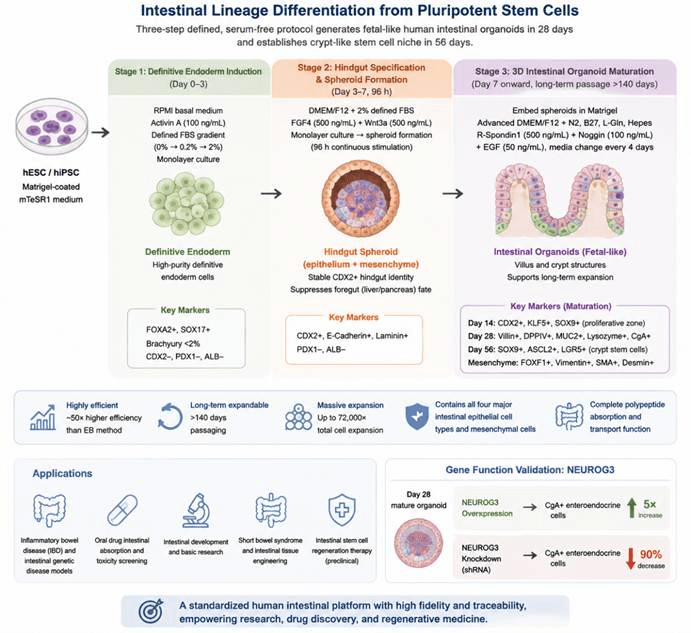
Differentiation process of pluripotent stem cells into intestinal lineage. hESC/iPSC are first induced into definitive endoderm by Activin A, followed by FGF4/Wnt3a-mediated posterior gut fate specification and hindgut spheroid formation. The hindgut spheroids are further cultured in a three-dimensional system supplemented with R-Spondin1, Noggin, and EGF to generate human intestinal organoids containing crypt–villus structures, intestinal stem cells, and multiple mature epithelial cell types. This system can be used for intestinal development studies, disease modeling, drug screening, and regenerative medicine research.
Differentiation Stage | Added Factors / Culture Conditions | Core Biological Function | Corresponding Cell Type / Embryonic Development Stage | Stage-Specific Identification Markers |
hPSC Maintenance (Starting Cells) | Matrigel-coated plates; feeder-free mTeSR1 medium; monolayer colony culture | Maintains the naive pluripotent state and preserves the potential for directed endoderm differentiation | Human embryonic stem cells / induced pluripotent stem cells (hES/hiPS) | Positive: OCT4, NANOG, SOX2Negative: FOXA2, SOX17, CDX2 and other endoderm markers |
Stage 1: Definitive Endoderm Induction (Day 0–3) | RPMI basal medium with defined fetal bovine serum gradient (0% → 0.2% → 2%); 100 ng/mL Activin A; monolayer adherent culture | Mimics embryonic gastrulation and efficiently induces highly purified definitive endoderm while suppressing mesodermal and ectodermal contamination | Embryonic definitive endoderm (upstream progenitor of intestinal development) | Strong positive: FOXA2, SOX17Mesoderm marker Brachyury <2%Negative: CDX2, PDX1, ALB (anterior/posterior gut markers) |
Stage 2: Hindgut Specification + Hindgut Spheroid Formation (Day 3–7, continuous 96 h stimulation) | DMEM/F12 + 2% defined fetal bovine serum; 500 ng/mL FGF4 + 500 ng/mL Wnt3a; monolayer culture with spontaneous formation of suspended three-dimensional spheroids | FGF4 promotes mesenchymal expansion and epithelial tube morphogenesis; Wnt3a cooperates with FGF4 to permanently suppress foregut (liver/pancreas) lineage and stabilize CDX2⁺ hindgut fate. Continuous 96 h stimulation ensures stable posterior gut specification | Embryonic hindgut epithelium + hindgut mesenchymal mixed three-dimensional spheroids (equivalent to mouse E8.5 hindgut tissue) | Broad positive: CDX2 (epithelial + mesenchymal), E-Cadherin, LamininCompletely negative: PDX1, ALB (foregut markers) |
Stage 3: Three-Dimensional Intestinal Organoid Maturation (Starting from Day 7, long-term expansion >140 days) | Hindgut spheroids embedded in Matrigel; Advanced DMEM/F12 supplemented with L-Gln, HEPES, N2, and B27; continuous addition of 500 ng/mL R-Spondin1, 100 ng/mL Noggin, and 50 ng/mL EGF; medium replacement every 4 days | The 3D extracellular matrix environment drives intestinal tissue maturation, gradually forming villus-like structures and crypt stem cell niches. It supports differentiation of multiple intestinal epithelial subtypes and intestinal mesenchymal cells, establishing an intestinal stem cell microenvironment for long-term expansion | Human fetal intestinal organoids (mimicking E12.5–E16.5 fetal intestine) | Early organoids (Day 14): CDX2, KLF5, widespread SOX9 (proliferative zone)Mature epithelium (Day 28): Villin, DPPIV (absorptive enterocytes), MUC2 (goblet cells), Lysozyme (Paneth cells), CgA (enteroendocrine cells)Stem cell niche (Day 56): SOX9, ASCL2, LGR5 (crypt-specific markers)Mesenchymal markers: FOXF1, Vimentin, SMA, Desmin |
Gene Function Validation (NEUROG3 Regulation Experiment) | Mature organoids at Day 28 were subjected to NEUROG3 overexpression or NEUROG3 shRNA knockdown | Validates NEUROG3 as an essential master transcription factor required for human intestinal enteroendocrine cell differentiation | Mature intestinal epithelial endocrine progenitor cells | Overexpression group: CgA⁺ enteroendocrine cells increased 5-foldKnockdown group: CgA⁺ cells decreased by 90% |