
info@ucallmlabs.com
Chronic kidney disease and end-stage renal failure affect a large global patient population, while the shortage of kidney organ donors remains a major challenge. Dialysis therapy is associated with high medical costs and reduced quality of life, making renal regenerative cell therapy and in vitro kidney disease models promising alternative approaches for research and development. All renal cell types, including renal tubule cells, glomerular cells, adrenal cells, and gonadal cells, originate from embryonic intermediate mesoderm (IM). In 2013, the Yamanaka laboratory at Kyoto University published a landmark induction technology in Nature Communications, establishing a serum-free, scalable, two-step monolayer single-cell directed differentiation protocol. By precisely controlling Activin A, Wnt/CHIR, and BMP7 signaling pathways in a temporal manner, this system first induces homogeneous mesendoderm and subsequently promotes efficient specification into intermediate mesoderm. Under optimized conditions, the induction efficiency of OSR1-positive IM cells reaches up to 90%, significantly outperforming traditional embryoid body-based and colony-based differentiation methods. This protocol is compatible with both H9 human embryonic stem cells (hESCs) and multiple patient-derived hiPSC lines.
The isolated OSR1-positive intermediate mesoderm cells retain broad multilineage differentiation potential and can be further induced in vitro into functional cell types including proximal tubular cells, glomerular podocytes, ureteric bud cells, adrenal cortical cells, and gonadal lineage cells. Following transplantation into immunodeficient mice, these cells do not form teratomas, and co-culture with embryonic kidneys enables the assembly of polarized functional renal tubule structures, demonstrating their safety and regenerative potential. The entire differentiation process is based on a two-dimensional monolayer culture system, making it suitable for high-throughput drug screening. The serum-free formulation also meets the requirements for cell therapy translational applications. Based on this technology, a standardized human intermediate mesoderm cell platform has been established for applications including hereditary kidney disease modeling, high-throughput nephrotoxicity evaluation of new drugs, renal tissue engineering seed cell preparation, and fundamental studies of human kidney embryonic development, providing an efficient, stable, and traceable human cell resource platform for renal regenerative medicine.
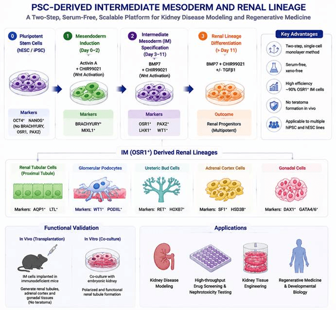
The process of differentiation of pluripotent stem cells into the renal lineage. hESC/iPSC undergo stepwise induction with Activin A, CHIR99021, and BMP7, sequentially transitioning through the mesendoderm and intermediate mesoderm (OSR1⁺ IM) stages, and further generating kidney-related cell types such as renal tubular cells, podocytes, and ureteric bud cells. This serum-free and scalable differentiation system provides a standardized human cell platform for kidney disease modeling, drug screening, and renal regeneration research.
Differentiation Stage | Added Factors / Culture Conditions | Core Biological Function | Corresponding Cell Type / Developmental Stage | Stage-Specific Identification Markers |
|---|---|---|---|---|
Human pluripotent stem cell maintenance (hiPSC/hESC) | Feeder-based system: primate ESC culture medium + 4 ng/mL bFGF; single-cell pretreatment: Accutase dissociation, 10 μM Y27632 (ROCK inhibitor) to maintain cell survival; feeder-free system: Matrigel-coated plates with mTeSR1 medium | Maintains pluripotent state and provides homogeneous starting cells for differentiation; inhibits apoptosis during single-cell dissociation and improves plating survival | Undifferentiated human induced pluripotent stem cells / human embryonic stem cells (primitive epiblast cells) | Basal pluripotency marker expression; negative for BRACHYURY, OSR1, PAX2 mesoderm markers; negative: SOX1, PAX6 (ectoderm), SOX17 (primitive endoderm) |
Stage 1: Mesendoderm (ME) induction (Differentiation day 0–2, single-cell monolayer method) | Basal medium: DMEM/F12 + 2% FBS (B27 supplement for serum-free system); key signals: 100 ng/mL Activin A + 3 μM CHIR99021 (Wnt3a substitute); matrix: Type I collagen-coated plates | Activates TGF-β/Activin and canonical Wnt signaling pathways, directing pluripotent cells toward mesendoderm fate; suppresses ectodermal and definitive endoderm differentiation | Embryonic mesendoderm (primitive streak cells, upstream renal developmental progenitors) | Highly expressed: BRACHYURY, MIXL1, GSC; very low/negative: OSR1, PAX2, SOX1, PAX6, SOX17 |
Stage 2: Intermediate mesoderm (IM) specification (Differentiation day 3–11, peak at D11) | Basal medium: DMEM/F12 + 10% KSR (B27 replacement for serum-free conditions); continuous signals: 100 ng/mL BMP7 + 3 μM CHIR99021; continuous monolayer culture with medium replacement every 2 days; control: Noggin/Frizzled-Fc strongly inhibits IM generation | BMP7 and Wnt signaling synergistically drive mesendoderm toward intermediate mesoderm differentiation; OSR1 functions as a key regulator of IM specification and efficiently enriches nephrogenic progenitors | Early intermediate mesoderm (nephric progenitors, common origin of kidney, adrenal gland, and gonad) | Positive markers: OSR1 (GFP reporter signal), PAX2, LIM1, WT1, CITED2, EYA1, SALL1; low expression of lateral plate mesoderm / hematopoietic marker CD41 |
Stage 3: IM downstream renal lineage maturation (additional 7-day culture after OSR1⁺ sorting) | Maintain 100 ng/mL BMP7 + 3 μM CHIR99021; addition of 10 ng/mL TGFβ1 promotes renal tubular epithelial formation | Drives intermediate mesoderm differentiation into renal tubules, glomerular cells, ureteric bud cells, adrenal cortical cells, and gonadal lineage cells | Metanephric mesenchyme, ureteric bud, proximal tubular cells, glomerular podocytes, adrenal/gonadal progenitor cells | 1. Ureteric bud: RET, SALL4, HOXB7, DBA lectin; 2. Metanephric mesenchyme: SIX2, HOXD11; 3. Proximal tubule: AQP1, LTL lectin; 4. Glomerular podocytes: Podocalyxin, PNA lectin; 5. Adrenal/gonadal lineage: GATA4, GATA6, HSD3β, SF1, DAX1; epithelial markers: E-Cadherin, cytokeratin |
In vivo transplantation validation | Flow-sorted D11 OSR1-GFP⁺ IM cells embedded in collagen sponge and implanted into the epididymal fat pad of immunodeficient NOD-scid mice | Validates in vivo differentiation potential of IM cells and confirms absence of teratoma formation, generating only IM-derived tissues | Mature renal tubular cells, adrenal cortical cells, and gonadal cells in vivo | Human cell markers + AQP1, LTL, DBA, E-Cadherin; absence of three-germ-layer teratoma tissues |
Mouse PSC-specific differentiation supplementation | Same two-step basic factors as human system; additional retinoic acid (RA) added during Stage 2 to enhance mouse Osr1 expression | Adapts the system for mouse iPS/ES cells and improves efficiency of mouse intermediate mesoderm induction | Mouse-derived intermediate mesoderm progenitor cells | Mouse homologous markers: mOsr1, Pax2, Lim1 |
Traditional control 1: EB three-dimensional embryoid body method | Embryoid bodies generated in low-attachment plates without matrix; same two-step induction factors; no ROCK inhibitor | Induces spontaneous mixed differentiation; IM induction efficiency only reaches 46.5% with high batch variability | Mixed three-germ-layer cell populations with limited intermediate mesoderm generation | OSR1-positive cell proportion significantly lower than single-cell monolayer method |
Traditional control 2: Undissociated colony-based monolayer method | hPSC colonies plated without Accutase single-cell dissociation | Uneven differentiation within colonies; IM induction efficiency only around 15% | Heterogeneous multi-lineage cell populations with low-abundance IM cells | Low OSR1 expression and high proportion of unwanted differentiated cells |