
info@mail.ucallm.com
1. Overview
Pseudorabies virus (PRV), also known as Suid herpesvirus 1 (SuHV-1), is a member of the genus Varicellovirus within the subfamily Alphaherpesvirinae of the family Herpesviridae. It is the causative agent of Aujeszky's disease in pigs. It is an enveloped, linear double-stranded DNA virus with a virion approximately 200–250 nm in diameter.
PRV was first described in 1902 by the Hungarian veterinarian Aujeszky. Pigs are the only natural host and reservoir of PRV, but the virus has an unusually broad host range, infecting nearly all mammals, including cattle, sheep, dogs, cats, mink, foxes, wolves, and rhesus macaques. In non-natural hosts, PRV infection is typically fatal, with fatality rates frequently reaching 100%. Since 2017, China has reported 31 laboratory-confirmed human cases of PRV infection, presenting with severe encephalitis, ocular infection, and neurological sequelae.
The PRV genome is a linear double-stranded DNA molecule of approximately 140–150 kb, encoding at least 70 different proteins. Since 2011, highly virulent PRV variants have emerged in China, against which traditional Bartha vaccines fail to provide sufficient cross-protection, leading to large-scale outbreaks in swine herds. Currently, no human PRV vaccine is licensed worldwide; swine vaccines remain the primary means of control.
2. Background Information
2.1 What is Pseudorabies Virus?
PRV is an enveloped, linear double-stranded DNA virus belonging to the genus Varicellovirus within the subfamily Alphaherpesvirinae of the family Herpesviridae. The virion consists of a core, capsid, tegument, and envelope, approximately 200–250 nm in diameter. The PRV genome is a linear double-stranded DNA molecule of approximately 140–150 kb, comprising two components: a long unique region (UL) and a short unique region (US), with the US region flanked by internal inverted repeats (IR) and terminal inverted repeats (TR). The genome encodes at least 70 proteins, which can be divided into three groups: (1) envelope glycoproteins that mediate virus entry and spread; (2) virus-encoded enzymes involved in DNA metabolism and/or phosphorylation; and (3) proteins involved in virion assembly.
Genomic organization of Mojiang paramyxovirus (PMID: 24865545)
2.2 PRV Genome & Classification
PRV belongs to the genus Varicellovirus within the subfamily Alphaherpesvirinae of the family Herpesviridae. Its genome is a linear double-stranded DNA of approximately 140–150 kb. PRV exhibits the hallmark characteristics of alphaherpesviruses: it can establish latent infection in sensory ganglia and can be reactivated by stressors or pathogens.
A 2026 metaviromic study analyzing pig samples from 17 provinces in China confirmed that variant genotype II PRV strains are the predominant circulating lineage in China.
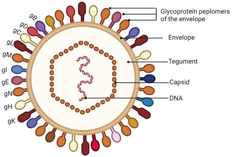
Schematic diagram of the PRV virion. (PMID: 36016260)
2.3 What Are PRV Proteins?
PRV encodes at least 70 proteins, among which the envelope glycoproteins play key roles in viral entry and immune responses.
Protein | Full name | Main function |
gB | Glycoprotein B | Viral entry (fusion); cell-cell spread; essential for virus viability |
gC | Glycoprotein C | Viral attachment; binds heparan sulfate |
gD | Glycoprotein D | Receptor binding (Nectin-1); essential for viral entry |
gE | Glycoprotein E | Viral spread in the nervous system; virulence factor |
gI | Glycoprotein I | Forms complex with gE; viral spread |
gH/gL | Glycoprotein H/L | Membrane fusion complex components |
gK | Glycoprotein K | Viral egress (secondary envelopment) |
TK | Thymidine kinase | Viral replication in neurons; virulence factor |
RR | Ribonucleotide reductase | DNA replication; virulence factor |
gB, gD, gH, and gL are essential for virus viability; PRV lacking these glycoproteins cannot penetrate host cells. gC, gE, gI, and gG are not necessary for virus replication, but gE-negative PRV has a greatly reduced capacity to infect second- or third-order neurons. Mutations in TK and RR genes significantly reduce viral replication and virulence in mice and pigs.
2.4 Mechanism of Cell Entry and Replication
2.4.1 Viral Attachment and Receptor Recognition
PRV infection begins with the binding of viral gC glycoprotein to heparan sulfate proteoglycans (HSPGs) on the host cell surface, mediating initial attachment. Subsequently, gD glycoprotein specifically interacts with the host cell receptor Nectin-1, a critical step for PRV entry. Additionally, Neuropilin-1 (NRP1) has been identified as an attachment and entry receptor for PRV. gD binding to Nectin-1 triggers activation of the PI3K/Akt signaling pathway, enhancing endocytosis and viral gene expression.
2.4.2 Viral Entry
PRV initiates entry through gD-Nectin-1 interaction. Following gD-receptor binding, the fusion complex composed of gB, gH, and gL mediates fusion of the viral envelope with the host cell membrane, releasing the viral nucleocapsid into the cytoplasm. PRV can infect multiple cell types, including porcine kidney cells (PK15), human neuroblastoma cells (SK-N-SH), and human embryonic kidney cells (HEK293T), demonstrating cross-species infectivity.
2.4.3 Viral RNA Replication
After the PRV genomic DNA enters the nucleus, it replicates using host cell machinery. Seven virus-encoded proteins are essential for viral DNA replication. TK and RR genes participate in DNA metabolism and support efficient viral replication in neurons.
2.4.4 Viral Assembly and Release
Viral DNA is packaged into capsids in the nucleus; capsids acquire a primary envelope by budding, then fuse with Golgi-derived vesicles to acquire the final envelope. gK participates in viral egress. Mature virions are transported via vesicles to the cell membrane and released.
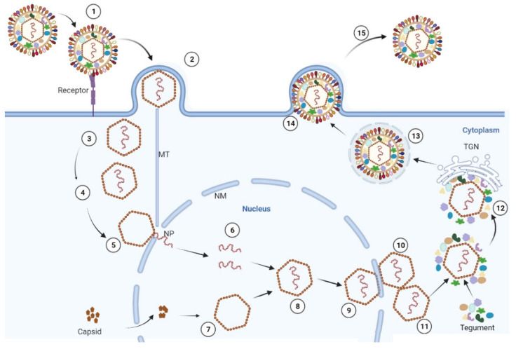
The replication cycle of PRV. (PMID: 36016260)
2.5 Symptoms and Treatment
2.5.1 Clinical Symptoms
Clinical manifestations in pigs vary depending on age, strain virulence, and immune status, including respiratory distress, neurological dysfunction, reproductive failure, and even death. Newborn piglets often develop acute encephalomyelitis, and pregnant sows may experience abortion, stillbirth, and mummified fetuses.
Human cases of PRV infection have been increasingly reported in China since 2017, with 31 laboratory-confirmed cases. Patients present with fever, visual impairment, headache, acute encephalitis, and permanent neurological sequelae. To date, no human-to-human transmission of PRV has been reported.
2.5.2 Antiviral Treatment
Currently, no specific antiviral therapy is approved for PRV. Treatment is primarily supportive. For human infection cases, screening and development of effective antiviral agents or immunotherapies should be prioritized.
2.6 Targets for Intervention
2.6.1 Viral Targets
Target | Protein | Biological Role |
gD | Glycoprotein D | Receptor binding |
gC | Glycoprotein C | Heparan sulfate binding |
DNA polymerase | DNA polymerase | DNA replication |
TK | Thymidine kinase | Viral replication |
gE | Glycoprotein E | Neural spread |
2.6.2 Host Receptors & Entry Factors
Host Target | Type | Role in Infection |
Nectin-1 | Receptor | Primary entry receptor |
NRP1 | Receptor | Attachment and entry receptor |
Heparan sulfate | Attachment factor | Initial attachment |
PILRα/PILRβ | Receptor | Possibly involved in entry |
2.6.3 Host Signaling Pathways
Pathway | Key Targets | Biological Role |
PI3K/Akt pathway | PI3K, Akt | Triggered by gD-receptor binding; enhances endocytosis and gene expression |
cGAS-STING pathway | cGAS, STING | Antiviral innate immunity |
JAK/STAT pathway | JAK1, STAT1 | IFN signaling, antagonized by TK protein |
NF-κB/MLKL pathway | NF-κB, MLKL | Necroptosis-mediated neuroinflammation |
2.7 Vaccine Types and Development Progress
2.7.1 Traditional Attenuated Live Vaccine (Bartha Strain)
Bartha-K61 is the most widely used PRV attenuated live vaccine globally, but it has limited protective efficacy against PRV variants that have emerged since 2011.
2.7.2 Genetically Engineered Deletion Vaccine
PRV-delgE/gI/TK is a triple-gene-deleted attenuated live vaccine. A 2026 study published in Microbiology Spectrum demonstrated that this vaccine preserves blood-brain barrier integrity and limits CNS injury following intracerebral PRV challenge.
2.7.3 Molecular Adjuvant-Enhanced Vaccine
In 2026, researchers constructed a TK/gE double-deleted recombinant PRV vaccine expressing CCL3L1 (HNX-ΔTK/ΔgE-CCL3L1). As a molecular adjuvant, CCL3L1 enhanced humoral immunity by activating the dendritic cell-T follicular helper cell-germinal center B cell axis, providing stronger protection than the traditional double-deletion vaccine.
2.7.4 Nanoparticle Vaccine
A 2026 study published in Vaccine designed a tetrameric antigen targeting gD and constructed a nanoparticle vaccine by coupling fluorinated polyethylenimine (FPEI) with λ-carrageenan (λ-CGN). The vaccine activated TLR4- and IRF3-mediated signaling pathways, inducing high levels of neutralizing antibodies and a Th1-biased cellular immune response in mice.
2.7.5 mRNA Vaccine
A 2025 study published in Journal of Virology developed a lipid nanoparticle-encapsulated gD mRNA vaccine (mRNA-LNPs) , evaluated in murine and porcine models. The vaccine induced potent neutralizing antibody responses and complete protection against lethal PRV variant challenge in mice. In piglets, immunization induced rapid humoral immune responses, significantly reduced viral loads in tissues and viral shedding post-challenge, and alleviated histopathological damage.
2.7.6 Subunit Vaccine
A 2026 study published in PubMed developed a CHO cell-expressed PRV gD subunit vaccine, which induced robust humoral immune responses and provided complete protection against lethal PRV variant challenge in mice.
2.8 Drugs
2.8.1 Virus-Targeted Drugs
Target/mechanism | Drug/candidate | Mechanism | Stage |
DNA polymerase | C2 (quinolone derivative) | up to 99% inhibition in vivo | Mouse model |
gD | binder-3 (mini-binder protein) | IC50= 2.41 nM | In vitro & mouse model |
gD + Nectin-1 | 3F7 mAb + Nectin-1 mAb | Dual-targeting strategy; synergistic enhancement of neutralization by 0.9–25.8% | In vitro |
gC-heparan sulfate | Aminoquinoline surfen | Blocks gC binding to heparan sulfate | In vitro & mouse model |
Host STING pathway | ST26 | STING agonist; host-directed antiviral | In vitro & mouse model |
PI3K-AKT/NF-κB pathway | Esculetin (coumarin) | Inhibits PI3K-AKT/NF-κB pathway activation | In vitro & in vivo |
Nectin-1 | B1 (mini-binder-derived small molecule) | IC50 = 1.2 nM | Mouse model |
2.9 Recent Research
2.9.1 Molecular Epidemiology and Surveillance
A 2025 systematic review published in Discover Nano comprehensively reviewed recent advances in the PRV genome, epidemiology, vaccine research, and molecular detection methods. A 2026 metaviromic study confirmed that variant genotype II PRV is the predominant circulating lineage in China, with PoCMV and PRV detected in all 17 sampled provinces. Furthermore, PRV is widely established in wild swine populations in Europe and North America, forming stable endemic cycles.
2.9.2 Latency and Reactivation Mechanisms
A 2025 study published in Science Advances revealed that pathogenic bacteria selectively trigger the transition of PRV from latency to reactivation. The study found that extraintestinal pathogenic Escherichia coli (ExPEC) and Streptococcus suis can trigger PRV reactivation; bacterial infection upregulates CXCL1 expression, enhancing STAT3 phosphorylation and nuclear translocation, thereby promoting early viral gene expression. Additionally, curcumin maintains PRV latent infection and inhibits reactivation by suppressing EGFR expression and phosphorylation.
2.9.3 Virus-Host Interactions and Pathogenesis
A 2025 study published in Journal of Virology revealed that PRV induces ferroptosis through TfR1 activation and ferritinophagy. PRV infection upregulates TfR1 expression via HIF-1β and promotes its translocation to the cell membrane through Rab11a, while also activating ferritinophagy to degrade FTH1, leading to intracellular iron overload. A 2026 study published in PNAS further found that PRV triggers ferroptosis by activating ferritinophagy, leading to prostaglandin E2 (PGE2) -mediated neuroinflammation; the ferroptosis inhibitor deferoxamine effectively mitigates PRV-induced viral encephalitis.
2.9.4 Virus-Host Interactions and Pathogenesis
Significant breakthroughs in PRV diagnostics were made in 2025–2026:
l CRISPR-Cas12b system: An ultrasensitive, visual PRV detection method was developed, enabling detection via real-time fluorescence readout or visualisation with a portable blue light instrument.
l Extraction-free LAMP-CRISPR/Cas12a assay: Targets the PRV gG gene with a detection limit of 1.0 × 10⁻⁴ copies/μL, yielding results within one hour.
l One-pot RAA-CRISPR/Cas12a assay: Detection limits of 5×10⁰ copies/μL (recombinant plasmid) and 7.96×10⁻² TCID50 per reaction (virus), completed within 50 minutes.
2.9.5 Zoonotic Transmission and Public Health Risk
A 2026 review published in Lancet Microbe systematically assessed the evidence, risk assessment, and future directions for human PRV infection. Despite the concerning surge in suspected human PRV cases in China, the risk of human PRV infection remains very low, and human-to-human transmission has never been reported. The researchers call for the eradication of PRV from domestic pigs through enhanced vaccination, biosecurity, and policy support to mitigate this emerging public health threat.
3. Related Products & Services
l Recombinant Antigens & Receptors
l Antibodies
l Detection Kits
l Recombinant Protein Expression Services
l Antibody Development Services
4. Resources
5. References
1. Pomeranz, L. E., Reynolds, A. E., Hengartner, C. J. (2005). Molecular biology of pseudorabies virus: impact on neurovirology and veterinary medicine. Microbiology and molecular biology reviews : MMBR, 69(3), 462–500.
2. Klupp, B. G., Hengartner, C. J., Mettenleiter, T. C., et al. (2004). Complete, annotated sequence of the pseudorabies virus genome. Journal of virology, 78(1), 424–440.
3. Mettenleiter T. C. (2000). Aujeszky's disease (pseudorabies) virus: the virus and molecular pathogenesis--state of the art, June 1999. Veterinary research, 31(1), 99–115.
4. Connolly, S. A., Jackson, J. O., Jardetzky, T. S., et al. (2011). Fusing structure and function: a structural view of the herpesvirus entry machinery. Nature reviews. Microbiology, 9(5), 369–381.
5. Geraghty, R. J., Krummenacher, C., Cohen, G. H., et al. (1998). Entry of alphaherpesviruses mediated by poliovirus receptor-related protein 1 and poliovirus receptor. Science (New York, N.Y.), 280(5369), 1618–1620.
6. Zheng, H. H., Fu, P. F., Chen, H. Y., et al (2022). Pseudorabies Virus: From Pathogenesis to Prevention Strategies. Viruses, 14(8), 1638.
7. An, T. Q., Peng, J. M., Tian, Z. J., et al. (2013). Pseudorabies virus variant in Bartha-K61-vaccinated pigs, China, 2012. Emerging infectious diseases, 19(11), 1749–1755.
8. Luo, Y., Li, N., Cong, X., et al. (2014). Pathogenicity and genomic characterization of a pseudorabies virus variant isolated from Bartha-K61-vaccinated swine population in China. Veterinary microbiology, 174(1-2), 107–115.
9. Wang, C. H., Yuan, J., Qin, H. Y., et al. (2014). A novel gE-deleted pseudorabies virus (PRV) provides rapid and complete protection from lethal challenge with the PRV variant emerging in Bartha-K61-vaccinated swine population in China. Vaccine, 32(27), 3379–3385.
10. Tong, W., Liu, F., Zheng, H., et al. (2015). Emergence of a Pseudorabies virus variant with increased virulence to piglets. Veterinary microbiology, 181(3-4), 236–240.
11. Ai, J. W., Weng, S. S., Cheng, Q., et al. (2018). Human Endophthalmitis Caused By Pseudorabies Virus Infection, China, 2017. Emerging infectious diseases, 24(6), 1087–1090.
12. Hou, Y., Wang, Y., Zhang, Y., et al. (2022). Human Encephalitis Caused by Pseudorabies Virus in China: A Case Report and Systematic Review. Vector borne and zoonotic diseases (Larchmont, N.Y.), 22(7), 391–396.
13. Wang, G., Zha, Z., Huang, P., et al. (2022). Structures of pseudorabies virus capsids. Nature communications, 13(1), 1533.
14. Engel, E. A., Card, J. P., Enquist, L. W. (2023). Transneuronal Circuit Analysis with Pseudorabies Viruses. Current protocols, 3(7), e841.