
info@mail.ucallm.com
Cellular senescence is a fundamental aging mechanism characterized by stable cell-cycle arrest, altered chromatin remodeling, metabolic reprogramming, and senescence-associated secretory phenotype (SASP). Current research focuses on identifying molecular regulators that distinguish beneficial senescence responses from pathological accumulation during aging and disease.
We provide an end-to-end tools — from core signaling targets to validated functional assays — enabling you to dissect every critical node of the senescence research with confidence.
Key Target Highlights
Core Molecular Targets for Senescence Research
Biological Process | Major Signaling Molecules | Research Significance | Common Applications |
DNA Damage Response | γH2AX, ATM, ATR, 53BP1 | Detect persistent DNA damage signaling | Stress-induced senescence models |
Cell Cycle Arrest | p16 , p21 , p53, RB | Defines irreversible growth arrest | Aging, cancer, regeneration studies |
SASP Regulation | NF-κB, C/EBPβ, IL-6, IL-8 | Controls inflammatory phenotype | Inflammaging studies |
Chromatin Remodeling | Lamin B1, H3K27me3, SAHF | Senescence-associated nuclear remodeling | Epigenetic aging research |
Metabolic Regulation | mTOR, AMPK, mitochondrial ROS | Links metabolism and senescence | Nutrient sensing studies |
Autophagy Control | ULK1, LC3, p62, Beclin-1 | Regulates senescence-associated proteostasis | Aging and longevity research |
² Key research trend:
Modern senescence studies increasingly move beyond single markers toward multi-parameter characterization, integrating DDR activation, cell-cycle arrest, SASP secretion, and metabolic alterations.
Recommended Senescence Marker Strategy
Multi-Parameter Validation Framework
Detection Layer | Recommended Markers | Purpose |
Primary Senescence Identification | p16, p21, SA-β-gal | Establish senescence phenotype |
DDR Confirmation | γH2AX, 53BP1, phospho-ATM | Verify DNA damage activation |
Inflammatory Phenotype | IL-6, IL-8, TNF-α, MCP-1 | Characterize SASP activity |
Cell Proliferation Status | Ki-67, EdU incorporation | Confirm growth arrest |
Chromatin Alteration | Lamin B1, H3K9me3 | Detect nuclear remodeling |
Metabolic State | mTOR, AMPK, ROS, mitochondrial markers | Analyze senescence metabolism |
Core Senescence Validation Strategy
Strategy | Marker Combination | Scientific Purpose | Recommended Application |
Golden Pair A: Senescence Establishment | p16/p21 + γH2AX | Links cell-cycle arrest with DDR activation | Replicative & stress-induced senescence |
Golden Pair B: Senescence Function | p16/p21 + SASP markers | Differentiates senescence from reversible arrest | Aging and inflammation studies |
Golden Pair C: Senescence Mechanism | γH2AX + NF-κB/SASP | Connects DNA damage to inflammatory signaling | Disease-associated senescence |
Competitive Technology Landscape
Senescence Research Workflow
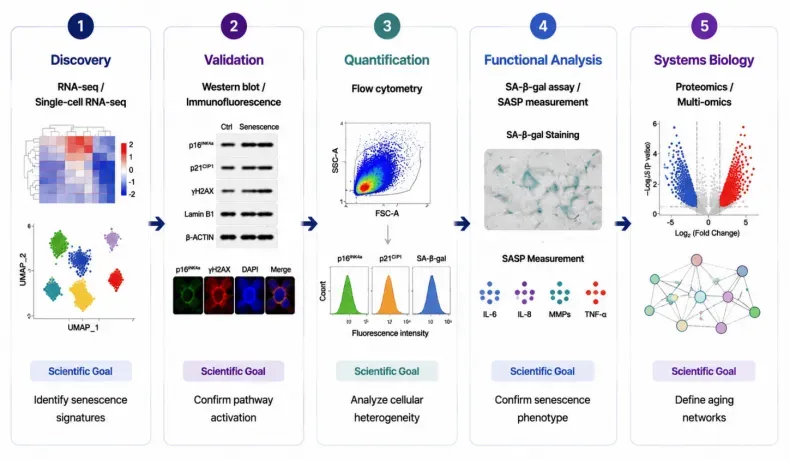
Cellular senescence research requires an integrated workflow combining molecular discovery, pathway validation, cellular quantification, functional characterization, and multi-omics analysis. This comprehensive strategy enables researchers to identify senescence signatures, define regulatory mechanisms, and uncover aging-associated biological networks.
Pathway Overview
Cellular senescence is a stress-responsive state characterized by irreversible cell-cycle arrest, altered chromatin organization, metabolic remodeling, and secretion of senescence-associated secretory phenotype (SASP) factors. Key regulatory networks, including p53–p21, p16INK4a–RB, DNA damage response (DDR), mTOR, and inflammatory signaling pathways, coordinate senescence induction and maintenance. Understanding senescence mechanisms provides critical insights into aging, tissue degeneration, cancer, and age-related diseases.
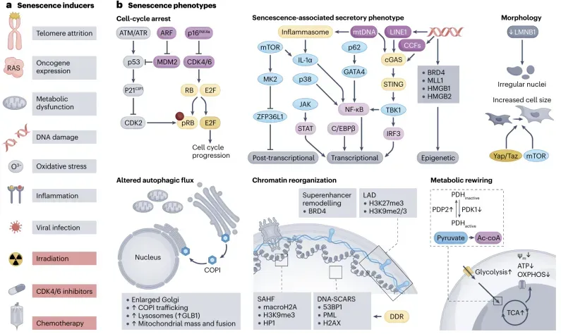
Different strategies to selectively eliminate senescent cells. (PMID: 39548312)
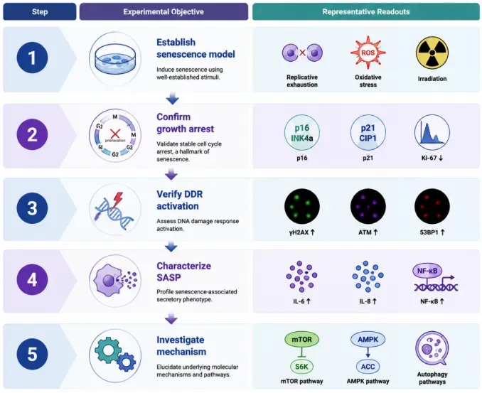
Recommended Experimental Validation Workflow
Step | Experimental Objective | Representative Readouts |
Step 1 | Establish senescence model | Replicative exhaustion, oxidative stress, irradiation |
Step 2 | Confirm growth arrest | p16, p21, Ki-67 |
Step 3 | Verify DDR activation | γH2AX, ATM, 53BP1 |
Step 4 | Characterize SASP | IL-6, IL-8, NF-κB |
Step 5 | Investigate mechanism | mTOR, AMPK, autophagy pathways |
Featured Research Application Examples
Research Area | Model System | Key Evaluation Parameters | Scientific Question |
Replicative Aging | Primary fibroblasts | p16/p21, telomere-associated DDR | How does cellular exhaustion occur? |
Therapy-Induced Senescence | Cancer models | DDR + SASP profiling | How does treatment trigger durable arrest? |
Tissue Aging | Animal/human tissues | Multiplex senescence markers | Where do senescent cells accumulate? |
Regenerative Biology | Stem cells | Senescence and inflammatory markers | How does aging impair regeneration? |
Frequently Asked Questions
Q1. Which markers should be used to confirm cellular senescence?
Cellular senescence is a complex and heterogeneous state that requires a combination of multiple complementary markers rather than a single indicator. Common senescence markers include p16INK4a, p21, and p53 for cell cycle arrest; SA-β-galactosidase activity for senescence-associated lysosomal changes; and γH2AX, 53BP1, and persistent DNA damage response (DDR) signaling for senescence-associated DNA damage. Additional markers such as Lamin B1 loss and SASP factors (IL-6, IL-8, MMPs) help define senescence phenotypes. Reliable senescence studies integrate cell cycle analysis, molecular markers, and functional validation to accurately characterize cellular senescence.
Q2. How can cellular senescence be distinguished from temporary cell cycle arrest or quiescence?
Senescence differs from reversible growth arrest through its stable proliferative arrest, altered cellular metabolism, and active secretory phenotype. Researchers should evaluate multiple parameters, including irreversible loss of proliferation capacity, increased p16/p21 signaling, elevated SA-β-gal activity, DNA damage accumulation, and SASP secretion. Appropriate controls, such as quiescent cells or serum-starved cells, are essential to distinguish true senescence from reversible growth inhibition.
Q3. What are the major signaling pathways regulating cellular senescence?
Cellular senescence is primarily controlled by the p53–p21 and p16–RB tumor suppressor pathways, which enforce stable cell cycle arrest. Additional regulatory networks include DNA damage response (DDR), mTOR signaling, AMPK metabolism pathways, NF-κB-mediated SASP regulation, and mitochondrial dysfunction-associated pathways. These signaling networks collectively regulate senescence induction, maintenance, inflammatory signaling, and cellular remodeling.
Q4. What are the common challenges and pitfalls in senescence research?
Major challenges include the lack of a universal senescence marker, biological heterogeneity across cell types, and difficulty distinguishing senescence from stress responses or apoptosis. Common pitfalls include relying only on SA-β-gal staining or a single protein marker. Best practices require combining senescence-associated markers (p16, p21, Lamin B1), SASP profiling, DNA damage assessment, and functional assays to achieve reliable conclusions.
Q5. How can senescence research be translated into aging mechanisms and therapeutic applications?
Senescence plays critical roles in aging, cancer, tissue regeneration, fibrosis, and chronic inflammation. Advanced studies integrate single-cell sequencing, spatial transcriptomics, proteomics, and SASP profiling to identify senescent cell populations and disease-associated signatures. Emerging therapeutic strategies, including senolytics and senomorphics, aim to selectively eliminate senescent cells or modulate their harmful secretory effects, providing potential approaches for age-related disease intervention.
Key References
1. Wang B., Han J., Elisseeff J.H., et al. (2024).The senescence-associated secretory phenotype and its physiological and pathological implications. Nature Reviews Molecular Cell Biology. 25:958–978.
2. Wu Z., Qu J., Liu G.H. (2024).Roles of chromatin and genome instability in cellular senescence and their relevance to ageing and related diseases.Nature Reviews Molecular Cell Biology. 25:979–1000.
3. Gorgoulis V., Adams P.D., Alimonti A., et al. (2019).Cellular Senescence: Defining a Path Forward.Cell. 179(4):813–827.
4. Hernandez-Segura A., Nehme J., Demaria M. (2018).Hallmarks of Cellular Senescence.Trends in Cell Biology. 28(6):436–453.
5. Di Micco R., Krizhanovsky V., Baker D., d’Adda di Fagagna F. (2021).Cellular senescence in ageing: from mechanisms to therapeutic opportunities.Nature Reviews Molecular Cell Biology. 22:75–95.
6. Kohli J., Wang B., Brandenburg S.M., et al. (2021).Algorithmic assessment of cellular senescence in experimental models.Nature Protocols. 16:2476–2499.
7. Ajoolabady A., Pratico D., Bahijri S., et al. (2025).Hallmarks of cellular senescence: biology, mechanisms, regulations.Experimental & Molecular Medicine. 57:1482–1491.
8. Ajoolabady A., Pratico D., Bahijri S., et al. (2025). Hallmarks and mechanisms of cellular senescence in aging and disease.Cell Death Discovery. 11:364.