info@mail.ucallm.com
Spatial Neuroscience
Decoding Biology In Situ, Connecting Proteins, Nucleic Acids, and Spatial Function
Neuroscience is increasingly moving beyond single-cell molecular characterization toward understanding where biological events occur and how cellular interactions shape brain function and disease progression.
Spatial neuroscience integrates multiplex imaging, molecular profiling, and cellular mapping technologies to reveal the complex organization of neural tissues, enabling researchers to investigate:
- Cellular composition and spatial distribution within brain regions
- Neural circuits and functional microenvironments
- Cell-cell communication networks
- Disease-associated cellular remodeling
- Regional vulnerability in neurodegenerative disorders
By combining validated cell-type markers with disease-related biomarkers, spatial profiling provides a powerful approach to connect molecular changes with anatomical context and biological function.
From Molecular Detection to Biological Insight
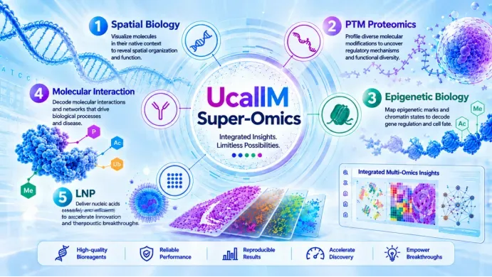
Super Omics™ integrates spatial biology, antibody-based detection, protein analysis, cellular and organoid models, nucleic acid delivery, and high-throughput technologies to support multidimensional investigation of biological systems. By connecting molecular detection, spatial context, cellular phenotypes, and functional analysis, Super Omics™ enables researchers to move from where molecules are → which cells are affected → how cellular interactions change → why disease phenotypes emerge.
Molecular Events → Cellular Phenotypes → Spatial Context → Functional Insight → Disease Mechanisms → Translational Discovery

Integrated Spatial Neuroscience Workflow
From Tissue Architecture to Mechanistic Discovery
1.1 Brain Tissue Architecture & Spatial Context
From Tissue Integrity to Spatially Resolved Biology
Key Biological Workflow
Brain Tissue Preservation——Tissue Architecture & Cellular Integrity——Regional and Cellular Mapping——Spatial Molecular Profiling
Research Focus
- Brain region-specific molecular characterization
- Preservation of tissue architecture and cellular morphology
- Cell-type identification and spatial distribution
- Regional differences in neuronal and glial composition
- Cellular neighborhood and tissue microenvironment analysis
- Spatially resolved investigation of disease-associated remodeling
Representative Markers
| Cellular Compartment | Representative Markers | Research Application |
|---|---|---|
| Neuronal Identity | NeuN/RBFOX3, MAP2, βIII-Tubulin/TUBB3 | Neuronal identification and spatial distribution |
| Microglia | IBA1/AIF1, TMEM119, P2RY12 | Microglial localization and state characterization |
| Astrocytes | GFAP, ALDH1L1, AQP4 | Astrocyte distribution and regional organization |
| Oligodendrocyte Lineage | OLIG2, SOX10, MBP, PLP1 | Oligodendrocyte lineage and myelin architecture |
| Neural Progenitors / Stem Cells | SOX2, Nestin | Neural progenitor localization where relevant |
| Vascular Compartment | CD31/PECAM1, EMCN, Collagen IV | Neurovascular architecture and BBB-associated spatial context |
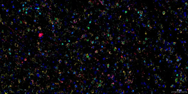
Neural Cell Atlas (Human Glioma FFPE)

1.2 Multiplex Antibody Panel Design
From Cell Identity to Disease-Associated Spatial Phenotypes
Key Biological Processes
Research Focus
- Neuronal identity and subtype composition
- Glial identity and activation states
- Synaptic architecture and organization
- Neurovascular and tissue microenvironment
- Disease-associated proteinopathies
- Spatial relationships between cells and pathological features
Representative Multiplex Panels
| Biological Component | Representative Markers | Primary Application |
|---|---|---|
| Neuronal Identity | NeuN/RBFOX3, MAP2, TUBB3 | Neuronal identification and distribution |
| Axonal Integrity | NF-L, NF-H, SMI-32 | Axonal architecture and neuronal injury |
| Synaptic Architecture | Synaptophysin, PSD95, Synapsin I | Synaptic density and pre-/postsynaptic organization |
| Excitatory Synapses | VGLUT1/SLC17A7, PSD95, GluA1 | Excitatory synapse characterization |
| Microglial Identity | IBA1/AIF1, TMEM119, P2RY12 | Microglial identification and homeostatic state |
| Microglial Activation / Phagocytosis | IBA1, CD68, MHC-II | Microglial activation and phagocytic response |
| Astrocyte Identity / Reactivity | ALDH1L1, GFAP, S100β, C3 | Astrocyte identity and reactive phenotypes |
| Oligodendrocyte / Myelin | OLIG2, SOX10, MBP, PLP1 | Oligodendrocyte lineage and myelin integrity |
| Neurodegenerative Pathology | Aβ, p-Tau, p-α-Syn, p-TDP-43 | Disease-associated proteinopathy |
| Neuroimmune Remodeling | C1q, C3, TREM2, APOE | Complement-associated and disease-associated remodeling |
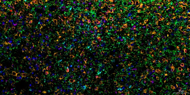
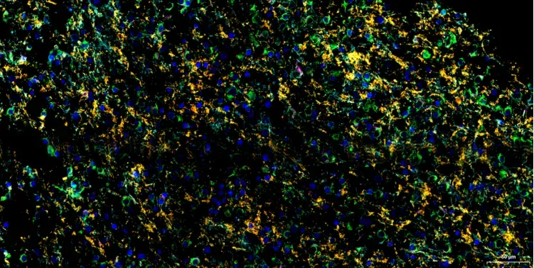
Neuronal & Synaptic Architecture (Human Glioma FFPE)

Rigorously Validated Antibody Panels
| Research Area | Target | Catalog No. | Product Name | Reactivity | Validated Applications |
|---|---|---|---|---|---|
| Neural Cell Atlas | OLIG2 | WR4617 | U-Blot® OLIG2 Rabbit mAb | Human, Mouse, Rat | WB, IHC, IF, ELISA |
| NeuN | WR4580 | U-Blot® NeuN Rabbit mAb | Human, Mouse, Rat | WB, IHC, IF, ELISA | |
| MAP2 | WR4578 | U-Blot® MAP2 Rabbit mAb | Human, Mouse, Rat | WB, IHC, IF, ELISA | |
| Iba1/AIF1 | WR4144 | U-Blot® Iba1 Rabbit mAb | Human, Mouse, Rat | WB, IHC, IF, IP, ELISA | |
| GFAP | WR4151 | U-Blot® GFAP Rabbit mAb | Human, Mouse, Rat | WB, IHC, IF, IP, ELISA | |
| DAPI | --- | --- | --- | --- | |
| Synaptophysin | WR4573 | U-Blot® Synaptophysin Rabbit mAb | Human, Mouse, Rat | WB, IHC, IF, ELISA | |
| SNAP25 | WR4968 | U-Blot® SNAP 25 Rabbit mAb | Human, Mouse, Rat | WB, IHC, IF, IP, ELISA | |
| PSD-95 | WR4266 | U-Blot® PSD95 Rabbit mAb | Human, Mouse, Rat | WB, IHC, IF, IP, ELISA | |
| NeuN / MAP2 / DAPI | WR4580 / WR4578 | U-Blot® NeuN / MAP2 Rabbit mAb | Human, Mouse, Rat | WB, IHC, IF, ELISA | |
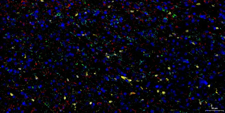
| Glial Cell Landscape | TMEM119 | WR5107 | U-Blot® TMEM119 Rabbit mAb | Mouse | WB, IHC, IF, ELISA |
| S100β/S100B | WR4271 | U-Blot® S100B Rabbit mAb | Human, Mouse, Rat | WB, IHC, IF, ELISA | |
| Iba1/AIF1 | WR4144 | U-Blot® Iba1 Rabbit mAb | Human, Mouse, Rat | WB, IHC, IF, IP, ELISA | |
| GFAP | WR4151 | U-Blot® GFAP Rabbit mAb | Human, Mouse, Rat | WB, IHC, IF, IP, ELISA | |
| AQP4 | WR5094 | U-Blot® AQP4 Rabbit mAb | Human, Mouse, Rat | WB, IHC, IF, IP, ELISA | |
| DAPI | --- | --- | --- | --- | |
| Neuronal Proteostasis & Protein Clearance | α-Synuclein | WR4587 | U-Blot® Synuclein-α Rabbit mAb | Human, Mouse, Rat, C.elegans | WB, IHC, IF, IP, ELISA |
| Tau | WR4576 | U-Blot® Tau Rabbit mAb | Human, Mouse, Rat | WB, IHC, IF, IP, ELISA |
1.3 Cellular Identification & Phenotyping
From Multiplex Imaging to Spatially Resolved Cellular Phenotypes
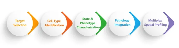
Key Analytical Workflow
Multiplex Image Acquisition——Image Processing & Quality Control——Cell&Nuclei Segmentation——Cell-Type Identification——Phenotypic Classification——Spatial Quantification & Cellular Mapping
Research Focus
- Neural Cell Identity
- Cellular State & Phenotype
- Cellular Function & Vulnerability
- Spatial Organization
- Disease-Associated Remodeling
| Cellular / Functional Category | Representative Markers | Primary Research Application |
|---|---|---|
| Pan-Neuronal Identity | NeuN/RBFOX3, MAP2, TUBB3 | Neuronal identification and spatial distribution |
| Neuronal / Axonal Integrity | NEFL, NEFH, SMI-32 | Axonal architecture and neuronal vulnerability |
| Excitatory Neurons | SLC17A7/VGLUT1, CAMKIIa | Excitatory neuronal populations |
| Inhibitory Neurons | GAD1, GAD2, GABA | GABAergic neuronal populations |
| Microglial Identity | IBA1/AIF1, TMEM119, P2RY12 | Microglial identification and homeostatic state |
| Microglial Activation / Phagocytosis | CD68, MHC-II, TREM2 | Activation, phagocytosis and disease-associated states |
| Disease-Associated Microglia | TREM2, APOE, LPL, CD68 | DAM-associated remodeling and lipid-response phenotypes |
| Astrocyte Identity | ALDH1L1, S100β, GFAP | Astrocyte identification and distribution |
| Reactive Astrocytes | GFAP, Vimentin, C3, SERPINA3N | Reactive and inflammatory astrocyte phenotypes |
| Oligodendrocyte Lineage | OLIG2, SOX10 | Oligodendrocyte lineage identification |
| Myelin Integrity | MBP, PLP1, CNP | Myelin organization and demyelination |
| Synaptic Organization | Synaptophysin, Synapsin I, PSD95, Homer1 | Synaptic density and pre-/postsynaptic organization |
| Neurovascular Compartment | CD31/PECAM1, EMCN, Collagen IV | Vascular architecture and neurovascular niche |
| Neurodegenerative Pathology | Aβ, p-Tau, p-α-Syn, p-TDP-43 | Disease-associated proteinopathy |
| Neuroimmune Remodeling | C1q, C3, TREM2, APOE | Complement and immune-associated remodeling |
| Cellular Stress / Injury | γH2AX, cleaved Caspase-3, HSP70 | DNA damage, apoptosis and cellular stress |
Glial Cell Landscape (Human Glioma FFPE)

1.4 Cell-Cell Interaction & Mechanistic Discovery
From Spatial Relationships to Disease Mechanisms
Key Analytical Workflow
Spatial Cell Mapping——Cellular Neighborhood Identification——Cell-Cell Proximity Analysis——Phenotypic Association——Disease Microenvironment Profiling——Mechanistic Hypothesis Generation
Research Focus
- Neuron-Microglia Crosstalk
- Neuron-Astrocyte Crosstalk
- Neuron-Oligodendrocyte Crosstalk
- Disease Microenvironment
Representative Spatial Neuroscience Research Panels
| Research Objective | Recommended Marker Panel | Biological Question |
|---|---|---|
| Brain Architecture Mapping | NeuN + MAP2 + GFAP + IBA1 | How are major neural cell populations spatially organized? |
| Neuroinflammation Profiling | IBA1 + TMEM119 + CD68 + GFAP + C3 | Where do reactive and inflammatory glial phenotypes emerge? |
| Syn |
Myelin & Axonal Integrity (Human Glioma FFPE)