
info@mail.ucallm.com
```
```
Apoptosis
1 Mechanism Overview
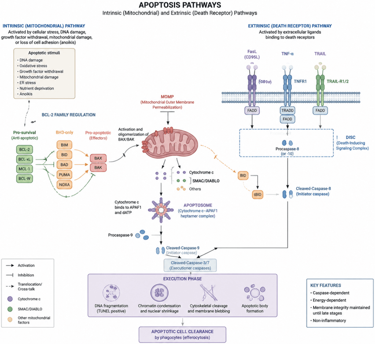
Apoptosis is a genetically regulated form of programmed cell death that is primarily mediated through the intrinsic mitochondrial pathway and the extrinsic death receptor pathway.The intrinsic pathway is mainly activated in response to intracellular stresses, including DNA damage, oxidative stress, endoplasmic reticulum stress, and mitochondrial dysfunction. This pathway is regulated by the BCL‑2 family proteins, which control mitochondrial outer membrane permeabilization (MOMP). Activation of the pro‑apoptotic proteins BAX/BAK induces the release of cytochrome c from mitochondria. Cytochrome c then interacts with Apaf‑1 to form the apoptosome, leading to the activation of Caspase‑9, which subsequently activates the executioner caspases Caspase‑3 and Caspase‑7 to initiate cell death.
The extrinsic pathway is triggered by the binding of death receptors, such as Fas, TNFR, and TRAIL receptors, to their corresponding ligands. This interaction promotes the recruitment of FADD and the formation of the death‑inducing signaling complex (DISC), resulting in the activation of Caspase‑8. Activated Caspase‑8 can directly activate executioner caspases or amplify mitochondrial apoptotic signaling through the cleavage of Bid into truncated Bid (tBid).
Both pathways ultimately converge on the activation of executioner caspases, mainly Caspase‑3, Caspase‑6, and Caspase‑7, which induce hallmark apoptotic events, including PARP cleavage, DNA fragmentation, chromatin condensation, cytoskeletal degradation, and apoptotic body formation. Meanwhile, regulatory factors such as p53, the inhibitor of apoptosis protein (IAP) family, and Smac/DIABLO collectively maintain precise control of apoptotic signaling.
Dysregulation of apoptosis, either through excessive activation or suppression, is closely associated with disease development. Apoptotic evasion is a critical mechanism underlying tumorigenesis and therapeutic resistance, whereas excessive apoptosis contributes to neurodegenerative diseases and tissue injury. Therefore, molecular markers including the BAX/BCL‑2 ratio, cytochrome c release, cleaved‑Caspase‑8/9, cleaved‑Caspase‑3, and cleaved‑PARP are widely used to evaluate the apoptotic process.
Apoptosis pathway schematic. The intrinsic mitochondrial pathway is triggered by intracellular stimuli such as DNA damage and oxidative stress. These signals are transmitted through the BCL‑2 family proteins, leading to the activation of BAX/BAK, which induces mitochondrial outer membrane permeabilization (MOMP). This results in the release of cytochrome c, followed by the formation of the apoptosome and activation of Caspase‑9.The extrinsic pathway is mediated by death receptors, including Fas, TNFR, and TRAIL receptors, which activate Caspase‑8 through the formation of the death‑inducing signaling complex (DISC). Both pathways ultimately converge on the activation of executioner Caspase‑3/7, leading to PARP cleavage, DNA fragmentation, chromatin condensation, cytoskeletal degradation, and apoptotic body formation, thereby completing the process of programmed cellular clearance.
2 Target List
| Mechanism Module | Target / Detection Marker | Type | Biological Function | Detection Significance |
|---|---|---|---|---|
| Overall Apoptotic Execution | Cleaved‑Caspase‑3 | Executioner caspase | Cleaves cellular structural proteins and induces cellular disassembly | Most widely used core marker for apoptosis confirmation |
| Cleaved‑Caspase‑7 | Executioner caspase | Cooperates with Caspase‑3 to execute apoptotic processes | Auxiliary marker for confirming apoptosis execution | |
| Cleaved‑PARP | Caspase substrate | Cleavage of PARP, a DNA repair protein | Terminal marker of apoptosis | |
| Intrinsic Mitochondrial Pathway | BAX | Pro‑apoptotic protein | Promotes mitochondrial outer membrane permeabilization (MOMP) | Determines activation of mitochondrial apoptosis |
| BAK | Pro‑apoptotic protein | Forms mitochondrial membrane pores together with BAX | Indicates occurrence of MOMP | |
| BCL‑2 | Anti‑apoptotic protein | Inhibits BAX/BAK activation | Evaluates anti‑apoptotic capacity | |
| BCL‑XL | Anti‑apoptotic protein | Maintains mitochondrial integrity | Evaluates inhibition of apoptosis | |
| MCL‑1 | Anti‑apoptotic protein | Suppresses mitochondrial apoptosis | Indicates cell survival signaling | |
| BH3‑only Pro‑apoptotic Initiators | BIM | Pro‑apoptotic factor | Activates BAX/BAK | Initiation signal of apoptosis |
| BID/tBID | Pro‑apoptotic factor | Connects death receptor signaling with mitochondrial pathway | Determines pathway crosstalk | |
| BAD | Pro‑apoptotic factor | Inhibits anti‑apoptotic function of BCL‑2 | Evaluates BCL‑2 family balance | |
| PUMA | p53 target gene | Promotes mitochondrial apoptosis | Marker of DNA damage‑induced apoptosis | |
| NOXA | BH3‑only protein | Inhibits MCL‑1 activity | Promotes MOMP induction | |
| Mitochondrial Release Events | Cytochrome c | Mitochondrial protein | Released into the cytoplasm after MOMP | Key evidence for mitochondrial apoptosis activation |
| SMAC/DIABLO | Mitochondrial protein | Inhibits IAP proteins and promotes caspase activation | Indicates amplification of apoptotic signaling | |
| Apoptosome Formation | APAF1 | Apoptosome component | Forms apoptosome complex with Cytochrome c | Confirms activation of intrinsic apoptotic pathway |
| Initiator Caspases | Cleaved‑Caspase‑9 | Initiator caspase | Activated by apoptosome formation | Marker of mitochondrial apoptotic pathway activation |
| Cleaved‑Caspase‑8 | Initiator caspase | Activated by death receptors and DISC complex | Marker of extrinsic apoptotic pathway activation | |
| Extrinsic Death Receptor Pathway | FAS/CD95 | Death receptor | Binds Fas ligand (FasL) to induce DISC formation | Determines Fas‑mediated apoptosis |
| TNFR1 | Death receptor | Mediates TNF‑α‑induced apoptotic signaling | Determines TNFR pathway activation | |
| TRAIL‑R1/R2 | Death receptor | Activates Caspase‑8 through TRAIL signaling | Determines TRAIL‑mediated apoptosis | |
| FADD | Adaptor protein | Recruits procaspase‑8 to form DISC complex | Core protein of extrinsic apoptotic pathway | |
| TRADD | Adaptor protein | Mediates TNFR1 signal transduction | Regulator of TNFR1‑mediated apoptosis | |
| Apoptosis Inhibitory Regulation | XIAP | IAP family protein | Inhibits Caspase‑3/7/9 activity | Determines mechanisms of apoptotic suppression |
| Cell‑level Validation | Annexin V | Membrane alteration marker | Detects phosphatidylserine externalization | Early apoptosis detection |
| PI (Propidium Iodide) | Membrane integrity marker | Distinguishes late apoptosis from necrosis | Used together with Annexin V analysis | |
| TUNEL | DNA damage marker | Detects DNA fragmentation | Validation of apoptosis in cells and tissues | |
| Hoechst/DAPI | Nuclear morphology marker | Detects chromatin condensation | Morphological confirmation of apoptosis |
3 Application Scheme
| Category | Marker Combination |
|---|---|
| Apoptosis occurrence | Cleaved‑Caspase‑3 + Cleaved‑PARP |
| Mitochondrial pathway (Intrinsic pathway) | BAX + BCL‑2 + Cytochrome c + Cleaved‑Caspase‑9 |
| Extrinsic pathway (Death receptor pathway) | FAS + FADD + Cleaved‑Caspase‑8 |
| Morphological validation | Annexin V + TUNEL |
4 References
- Yuan J, Ofengeim D. A guide to cell death pathways. Nat Rev Mol Cell Biol. 2024;25:379‑395. doi:10.1038/s41580‑023‑00689‑6.
- Czabotar PE, Garcia‑Saez AJ. Mechanisms of BCL‑2 family proteins in mitochondrial apoptosis. Nat Rev Mol Cell Biol. 2023;24:732‑748. doi:10.1038/s41580‑023‑00629‑4.
- Taylor RC, Cullen SP, Martin SJ. Apoptosis: controlled demolition at the cellular level. Nat Rev Mol Cell Biol. 2008;9:231‑241. doi:10.1038/nrm2312.
- Wang Y, et al. Cell death pathways: molecular mechanisms and therapeutic targets for cancer. MedComm. 2024;5:e693. doi:10.1002/mco2.693.