
info@mail.ucallm.com
```
```
Lysosome‑dependent cell death, LDCD
1 Mechanism Overview
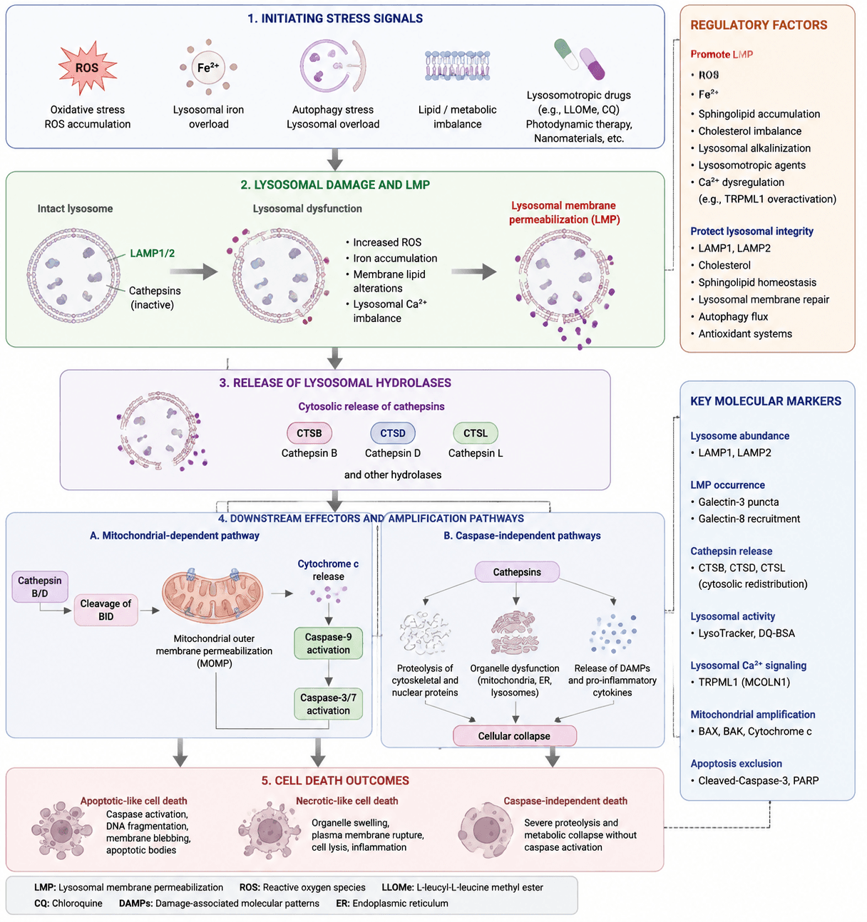
Lysosome‑dependent cell death (LDCD) is a regulated form of cell death driven by lysosomal dysfunction and lysosomal membrane permeabilization (LMP). Various cellular stressors, including oxidative stress (ROS accumulation), lysosomal iron overload, lipid metabolic disorders, autophagic stress, and lysosome‑targeting compound stimulation, can reduce lysosomal membrane stability and induce LMP.
Damaged lysosomes release large amounts of acidic hydrolases, particularly the cysteine proteases Cathepsin B (CTSB) and Cathepsin L (CTSL), as well as the aspartic protease Cathepsin D (CTSD), into the cytoplasm, thereby triggering downstream death signaling.
On one hand, Cathepsins can promote mitochondrial outer membrane permeabilization (MOMP) by cleaving the BH3‑only protein Bid, leading to cytochrome c release and activation of the caspase cascade, resulting in apoptosis‑like cell death. On the other hand, extensive Cathepsin‑mediated proteolysis can directly disrupt the cytoskeleton, nuclear proteins, and organelle homeostasis, inducing caspase‑independent cell death.
Furthermore, lysosomal damage can promote the release of damage‑associated molecular patterns (DAMPs) and activation of inflammatory signaling, thereby amplifying cellular injury responses.
Lysosome‑dependent cell death pathway schematic. Lysosome‑dependent cell death is initiated by lysosomal stress and membrane destabilization, leading to lysosomal membrane permeabilization (LMP)‑mediated release of cathepsins. Cathepsins subsequently induce mitochondrial dysfunction, proteolytic degradation, and activation of inflammatory signaling, ultimately resulting in apoptosis‑like, necrosis‑like, or caspase‑independent cell death.
2 Target List
| Mechanism Module | Target / Detection Marker | Full Name | Biological Function | Detection Method |
|---|---|---|---|---|
| Lysosomal Identification and Function | LAMP1 | Lysosome‑associated membrane glycoprotein 1 | Classical lysosomal membrane protein; evaluates lysosome abundance and localization | WB / IF / IHC |
| LAMP2 | Lysosome‑associated membrane glycoprotein 2 | Maintains lysosomal membrane stability and reflects lysosomal integrity | WB / IF | |
| LysoTracker | Lysosome‑specific acidic organelle fluorescent probe | Detects acidic lysosome abundance and acidification status | IF / Flow cytometry | |
| DQ‑BSA | Dye‑quenched bovine serum albumin | Evaluates lysosomal protein degradation capacity | Fluorescence assay | |
| Lysosomal Membrane Permeabilization (LMP) | Galectin‑3 puncta | Galectin‑3 puncta formation | Marker of damaged lysosomal membranes | IF microscopy |
| Galectin‑8 recruitment | Galectin‑8 recruitment to damaged lysosomes | Identifies damaged lysosomes | IF | |
| LAMP1 redistribution | Lysosome‑associated membrane glycoprotein 1 redistribution | Determines abnormalities in lysosomal membrane structure | IF | |
| Acridine orange release | Acridine orange release assay | Detects loss of lysosomal membrane integrity | Flow cytometry | |
| Cathepsin Release and Execution | CTSB | Cathepsin B | Lysosomal cysteine protease | WB / IF / Activity assay |
| CTSD | Cathepsin D | Lysosomal aspartic protease involved in protein degradation | WB / IF | |
| CTSL | Cathepsin L | Lysosomal cysteine protease involved in degradation of structural proteins | WB / IF | |
| Lysosomal Regulation and Homeostasis | MCOLN1 (TRPML1) | Mucolipin‑1 / Transient receptor potential mucolipin 1 | Lysosomal Ca²⁺ channel regulating calcium release | WB / IF |
| TFEB | Transcription factor EB | Master regulator of lysosome biogenesis and repair | WB / IF | |
| ATP6V1A / ATP6V1B2 | V‑type proton ATPase catalytic subunit A / subunit B2 | Maintains V‑ATPase function and lysosomal acidification | WB | |
| Oxidative Stress‑Induced Lysosomal Damage | ROS | Reactive oxygen species | Oxidative stress mediator | ROS probe assay |
| 4‑HNE | 4‑Hydroxynonenal | Lipid peroxidation product | WB / IF | |
| MDA | Malondialdehyde | Lipid oxidation end product | Biochemical assay |
3 Application Scheme
| Biological event | Marker |
|---|---|
| Lysosome abundance | LAMP1, LAMP2 |
| LMP | Galectin‑3 puncta |
| Lysosomal leakage | CTSB/CTSD redistribution |
| Lysosomal activity | LysoTracker, DQ‑BSA |
| Lysosomal Ca²⁺ | TRPML1 (MCOLN1) |
| Mitochondrial damage | BAX, BAK, Cytochrome c |
| Apoptosis exclusion | Cleaved‑Caspase3, PARP |
4 References
- Boya P, Kroemer G. Lysosomal membrane permeabilization in cell death. Oncogene. 2008;27(50):6434‑6451. doi:10.1038/onc.2008.310.
- Repnik U, Stoka V, Turk V, Turk B. Lysosomal cell death at a glance. Journal of Cell Science. 2014;127(1):3‑10. doi:10.1242/jcs.091181.
- Wang F, Gómez‑Sintes R, Boya P. Lysosomal membrane permeabilization and cell death. Traffic. 2018;19(12):918‑931. doi:10.1111/tra.12613.