
info@mail.ucallm.com
```
```
Autophagy‑dependent cell death, ADCD
1 Mechanism Overview
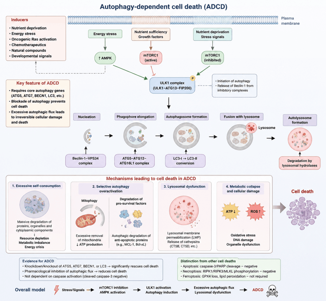
Autophagy‑dependent cell death (ADCD) is a form of programmed cell death that is executed through the core molecular machinery of autophagy. ADCD is typically induced by various intracellular and extracellular stressors, including nutrient deprivation, metabolic stress, alterations in developmental signals, pharmacological stimulation, and aberrant oncogene activation. Its core regulatory mechanism is closely associated with the mTOR (mechanistic target of rapamycin) signaling pathway.
Under nutrient‑rich conditions, mTORC1 remains activated and suppresses autophagy initiation by inhibiting the ULK1 complex. However, under conditions of nutrient deprivation or energy stress, mTORC1 activity is inhibited, resulting in the activation of the ULK1/ATG13/FIP200 complex, which initiates autophagosome formation. Subsequently, the Beclin‑1–VPS34 complex promotes phagophore nucleation, while the ATG5–ATG12–ATG16L complex mediates membrane elongation. The conversion of LC3‑I to LC3‑II facilitates autophagosome maturation, ultimately establishing a complete autophagic process.
During ADCD, sustained enhancement of autophagic flux serves as a critical driver of cell death. Unlike moderate autophagy that maintains cellular homeostasis, autophagy in ADCD is excessively activated, resulting in continuous degradation of large amounts of intracellular proteins, organelles, and cytoplasmic components. When autophagic activity exceeds the cellular compensatory capacity, excessive depletion of cellular resources occurs, leading to metabolic imbalance, insufficient energy supply, and structural damage, ultimately resulting in irreversible cell death. Therefore, “excessive self‑consumption” is considered a fundamental concept underlying ADCD.
The mechanism of ADCD can be summarized as follows: external or endogenous stress signals induce mTORC1 inhibition and ULK1 activation, initiating an ATG‑dependent autophagic program; subsequently, sustained autophagic flux promotes extensive degradation of cellular components, while selective autophagy eliminates essential cellular structures or survival factors and induces lysosomal stress and metabolic collapse; ultimately, irreversible cellular damage and cell death occur.
ADCD represents a novel form of programmed cell death distinct from classical death modalities such as apoptosis, necrosis, and ferroptosis. It plays important roles in developmental regulation, cancer therapy, and disease mechanism studies.
Autophagy‑dependent cell death pathway schematic. Stress stimuli (nutrient deprivation, energy stress, drugs, etc.) activate AMPK and inhibit mTORC1, thereby initiating the ULK1‑dependent autophagic program. Subsequently, ATG proteins mediate autophagosome formation and enhance autophagic flux. When autophagy exceeds the capacity required for cellular homeostasis maintenance, excessive self‑consumption, selective organelle degradation, lysosomal damage, and metabolic collapse ultimately lead to autophagy‑dependent cell death (ADCD).
2 Target List
| Mechanism Module | Target / Detection Marker | Full Name | Functional / Mechanistic Significance | Recommended Detection |
|---|---|---|---|---|
| Core Confirmation of ADCD | ATG5 | Autophagy‑related protein 5 | Essential protein for autophagosome formation; knockdown can block ADCD | WB / qPCR / KO or KD rescue experiments |
| ATG7 | Autophagy‑related protein 7 | Core autophagy execution factor involved in LC3 lipidation | WB / qPCR | |
| BECN1 (Beclin‑1) | Beclin‑1 autophagy‑related protein | Initiates autophagy and promotes PI3K complex formation | WB / IF | |
| LC3B (LC3‑I/LC3‑II) | Microtubule‑associated protein 1 light chain 3 beta | Classical marker of autophagosome formation and autophagic conversion | WB / IF | |
| ATG12, ATG16L1 | Autophagy‑related protein 12 / Autophagy‑related protein 16‑like 1 | Components of the ATG5–ATG12–ATG16 complex | WB | |
| Autophagy Initiation Regulation | p‑mTOR/mTOR | Phosphorylated mechanistic target of rapamycin / mechanistic target of rapamycin | mTORC1 inhibition promotes ADCD initiation | WB |
| ULK1/p‑ULK1 | Unc‑51 like autophagy activating kinase 1 | Core activation factor of the autophagy initiation complex | WB | |
| ATG13, FIP200/RB1CC1 | Autophagy‑related protein 13 / RB1‑inducible coiled‑coil protein 1 | Components of the ULK1 initiation complex | WB | |
| p‑AMPK/AMPK | Phosphorylated AMP‑activated protein kinase / AMP‑activated protein kinase | Energy stress sensor promoting autophagy activation | WB | |
| Autophagic Flux Assessment | p62/SQSTM1 | Sequestosome 1 | Autophagy substrate; decreased level indicates enhanced autophagic flux | WB / IF |
| LAMP1/LAMP2 | Lysosome‑associated membrane glycoprotein 1/2 | Markers of autophagosome–lysosome fusion | WB / IF | |
| CTSB (Cathepsin B) | Cathepsin B | Lysosomal hydrolase involved in cell death execution | WB / IF | |
| CTSD (Cathepsin D) | Cathepsin D | Lysosomal protein degradation function | WB | |
| Selective Autophagy / Mitophagy | PINK1 | PTEN‑induced kinase 1 | Recognition of damaged mitochondria | WB |
| Parkin/PARK2 | Parkin RBR E3 ubiquitin‑protein ligase | Promotes mitochondrial ubiquitination and clearance | WB | |
| BNIP3/NIX | BCL2 interacting protein 3 / BNIP3‑like protein | Receptor‑mediated mitophagy regulators | WB | |
| TOM20 | Translocase of outer mitochondrial membrane 20 | Indicator of mitochondrial content; decreases during excessive mitochondrial clearance | WB / IF | |
| COX IV | Cytochrome c oxidase subunit IV | Evaluation of mitochondrial quality | WB | |
| Lysosome‑Dependent Death Mechanisms | LAMP1 | Lysosome‑associated membrane glycoprotein 1 | Marker of autolysosome formation | WB / IF |
| TFEB | Transcription factor EB | Transcriptional regulator of lysosome biogenesis and autophagy | WB | |
| GBA1/GALC | Glucosylceramidase beta 1 / Galactosylceramidase | Lysosomal metabolism‑related factors involved in certain ADCD processes | WB | |
| Metabolic Depletion Mechanisms | ATP level | Cellular ATP level | Reflects excessive autophagy‑induced energy depletion | Biochemical assay |
| AMPK | AMP‑activated protein kinase | Regulator of energy stress response | WB | |
| HK2, GLUT1, LDHA | Hexokinase 2 / Glucose transporter 1 / Lactate dehydrogenase A | Indicators of glycolytic metabolic alterations | WB / qPCR | |
| Exclusion of Apoptosis | Cleaved‑Caspase‑3 | Cleaved caspase‑3 | Determines whether cell death is mediated by classical apoptosis | WB |
| Cleaved‑PARP | Cleaved poly(ADP‑ribose) polymerase | Execution marker of apoptosis | WB | |
| BAX/BCL‑2 | BCL2‑associated X protein / B‑cell lymphoma‑2 | Markers of mitochondrial apoptotic pathway | WB | |
| Exclusion of Ferroptosis | GPX4 | Glutathione peroxidase 4 | Ferroptosis inhibitory protein | WB |
| SLC7A11 | Solute carrier family 7 member 11 | Regulator of glutathione metabolism and cystine transport | WB | |
| ACSL4 | Acyl‑CoA synthetase long‑chain family member 4 | Ferroptosis‑promoting factor involved in PUFA metabolism | WB | |
| Exclusion of Necroptosis | RIPK1/RIPK3/p‑MLKL | Receptor‑interacting protein kinase 1 / 3 / phosphorylated mixed lineage kinase domain‑like protein | Markers used to exclude necroptotic signaling | WB |
3 Application Scheme
| Function | Markers |
|---|---|
| Autophagy initiation | ULK1, ATG13, BECN1 |
| Autophagosome formation | LC3‑I/II, ATG5, ATG7 |
| Autophagic flux | p62/SQSTM1, LAMP1 |
| Lysosomal function | LAMP1, Cathepsin B/D |
| mTOR regulation | p‑mTOR, p‑S6K, p‑ULK1 |
| Mitochondrial clearance (Mitophagy) | PINK1, Parkin, TOM20 |
| Exclusion of apoptosis | Caspase‑3, PARP |
| Cell death confirmation | ATG5/ATG7 knockdown rescue experiments |
4 References
- Denton D, Kumar S. Autophagy‑dependent cell death. Cell Death Differ. 2019;26(4):605‑616. doi:10.1038/s41418‑018‑0252‑y.
- Jung S, Jeong H, Yu SW. Autophagy as a decisive process for cell death. Exp Mol Med. 2020;52:921‑930. doi:10.1038/s12276‑020‑0455‑4.
- Huang X, Yan H, Xu Z, et al. The inducible role of autophagy in cell death: emerging evidence and future perspectives. Cell Commun Signal. 2025;23:151.